. 2005 May;79(10):6291-8.
doi: 10.1128/JVI.79.10.6291-6298.2005.

New antiviral pathway that mediates hepatitis C virus replicon interferon sensitivity through ADAR1

Affiliations

New antiviral pathway that mediates hepatitis C virus replicon interferon sensitivity through ADAR1

Deborah R Taylor et al. J Virol. 2005 May.

Abstract

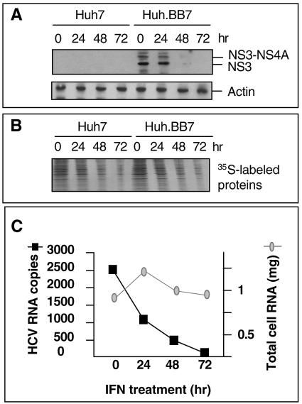
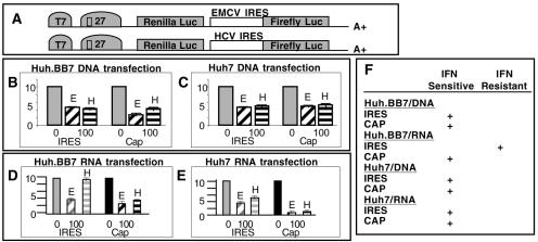
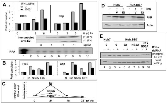
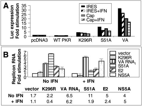
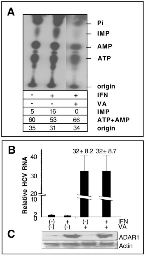
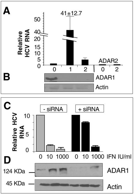
While many clinical hepatitis C virus (HCV) infections are resistant to alpha interferon (IFN-alpha) therapy, subgenomic in vitro self-replicating HCV RNAs (HCV replicons) are characterized by marked IFN-alpha sensitivity. IFN-alpha treatment of replicon-containing cells results in a rapid loss of viral RNA via translation inhibition through double-stranded RNA-activated protein kinase (PKR) and also through a new pathway involving RNA editing by an adenosine deaminase that acts on double-stranded RNA (ADAR1). More than 200 genes are induced by IFN-alpha, and yet only a few are attributed with an antiviral role. We show that inhibition of both PKR and ADAR1 by the addition of adenovirus-associated RNA stimulates replicon expression and reduces the amount of inosine recovered from RNA in replicon cells. Small inhibitory RNA, specific for ADAR1, stimulated the replicon 40-fold, indicating that ADAR1 has a role in limiting replication of the viral RNA. This is the first report of ADAR's involvement in a potent antiviral pathway and its action to specifically eliminate HCV RNA through adenosine to inosine editing. These results may explain successful HCV replicon clearance by IFN-alpha in vitro and may provide a promising new therapeutic strategy for HCV as well as other viral infections.


Figures

FIG. 1.
FIG. 1.
IFN-α action is replicon specific. (A) Immunoblot of HCV NS3 protein expression in lysates (25 μg) from parental Huh7 cells or replicon-containing cells (Huh.BB7) treated with IFN-α (100 IU/ml) for 24, 48, or 72 h. (B) Total cellular proteins from an equal number of [35S]methionine metabolically labeled cells after treatment with IFN-α (100 IU/ml) for 24, 48, or 72 h. (C) HCV RNA from 0.5 × 10⁶ cells was quantitated by Taqman RT-PCR using HCV standards. Total RNA from the same cells was measured by absorbance of UV (wavelength of 260 nm).
FIG. 2.
FIG. 2.
Cap-independent translation exhibits IFN-α resistance. (A) Bicistronic luciferase reporters expressing Renilla luciferase under control of a cap-dependent translation mechanism and firefly luciferase under the EMCV or HCV IRES. (B and C) Luc activity expressed as fold inhibition as a result of IFN-α treatment in cell lysates from Huh7 or Huh.BB7 cells transfected with bicistronic reporter plasmid DNA (2 μg) containing the EMCV (E) IRES or HCV (H) IRES constructs shown in panel A, expressed relative to the control (no IFN-α) (which was assigned a value of 10). (D and E) IFN-α (100 IU/ml) inhibition of Luc activity in cell lysates from equal numbers of Huh.BB7 or Huh7 cells transfected with bicistronic reporter plasmid RNA (synthesized in vitro using T7 RNA polymerase) containing the EMCV (E) or HCV (H) IRES. Luc assay results shown are representative of four or more experiments with transfections performed in duplicate and assayed for Luc activity in triplicate. Transfection efficiency was also monitored by RNase protection analysis (data not shown). Values are means ± standard deviations of the means (error bars). (F) Summary of data in panels B to E.
FIG. 3.
FIG. 3.
Inhibition of PKR stimulates translation but does not result in complete rescue of the replicon from the effects of IFN-α. (A) Luciferase activity measured in cell lysates from Huh.BB7 cells transfected with HCV IRES bicistronic reporter plasmid (2 μg) and E2 and/or pcDNA3 plasmids (10 μg total DNA/6-cm dish). Cells were untreated [(−) IFN] or treated with 1,500 IU/ml IFN-α [(+) IFN] for 18 h. The expression levels of transfected E2 are shown below the bars. Immunoblot of E2 from transfected cells. RNA was isolated from transfected cells, and Luc expression/transfection efficiency was monitored by RNase protection analysis (RPA) as described in Materials and Methods. (B) Luciferase activity in cells cotransfected with HCV IRES bicistronic reporter plasmid and E2 or NS5A (6 μg) with pcDNA3 (6 μg) or with both E2 and NS5A (E+N; 6 μg each). Luciferase activity is expressed as fold increase. Values are means ± standard deviations of the means (error bars). (C) Huh.BB7 cells transfected in triplicate with E2, NS5A, or pcDNA3 (vector). Cells were untreated (no IFN-α) or treated with 100 IU/ml IFN-α at 8 h posttransfection (72 h), 32 h posttransfection (48 h), or 56 h posttransfection (24 h) and were harvested at 80 h posttransfection. Replicon RNA was measured by Taqman analysis of HCV RNA relative to the level of GAPDH mRNA in 0.5 × 10⁶ cells (16). Values are means ± standard deviations of the means (error bars). (D) Cells transfected with pcDNA3 vector (V) or E2 pcDNA3 (E2), were treated with 1,000 IU/ml IFN-α for 18 h (+) and analyzed for PKR or actin expression by protein immunoblotting with polyclonal anti-PKR antiserum or antiactin antibodies. (E) Immunoblot detection of phosphorylated eIF-2α (phospho-eIF-2α) and total eIF-2α (20 μg total protein/lane) from cells transfected with E2 or NS5A or E2 plus NS5A and treated with 1,000 IU/ml IFN-α and poly(I · C) (20 μg/ml) for 18 h (+).
FIG. 4.
FIG. 4.
VA RNAI rescues the replicon from the effects of IFN-α. (A) Luciferase activity in Huh.BB7 cells cotransfected with HCV IRES bicistronic reporter and pcDNA3, wild-type PKR, PKR K296R, eIF-2α S51A, or VA RNAI. Cells were treated with 500 IU/ml IFN-α for 18 h. Luc activity is relative to the pcDNA3 control value, which was assigned a value of 1. (B) Taqman quantitation of HCV RNA from Huh.BB7 cells transfected with pcDNA3 (vector) or PKR inhibitors. Cells were untreated or treated with IFN-α at 500 IU/ml for 18 h (+). The histogram shows the fold increase in RNA detected from an equal number of inhibitor-transfected cells relative to vector-transfected cells, which was assigned a value of 1, relative to the amount of GAPDH mRNA. Values are means ± standard deviations of the means (error bars). Below the histogram are the actual RNA copy numbers (106) per reaction based on Taqman HCV RNA standards.
FIG. 5.
FIG. 5.
IFN-α-treated Huh.BB7 cells yield edited replicon RNA. Four regions within the HCV IRES of BB7 replicon that contain mutations are shown. The wild-type BB7 sequences are shown. The nucleotides in the wild-type BB7 sequence that were mutated are shown in bold type and underlined, and the nucleotides obtained after IFN-α treatment are shown below the sequence.
FIG. 6.
FIG. 6.
A-to-I RNA editing in replicon-containing cells. (A) Thin-layer chromatography of nuclease P1-digested RNA from Huh.BB7 cells grown in the presence of [α-32P]ATP with (+) and without (−) 100 IU/ml IFN-α in the absence (−) and presence (+) of transfected VA RNAI plasmid. Monophosphates were resolved on one TLC plate, although the rightmost lane was exposed longer. Radioactivity was quantitated by PhosphorImager analysis and is shown below the gel as a percentage of the total counts (IMP, ATP, AMP, and origin). (B) Replicon-containing cells were transfected with VA RNA and treated with IFN-α (+). HCV replicon RNA was monitored by Taqman analysis and was quantitated relative to the amount of GAPDH mRNA (16). Values are means ± standard deviations of the means (error bars). Transfections were performed in duplicate with one dish of cells used for Taqman analysis and the other used for immunoblot analysis (C) where cytoplasmic extracts (20 μg) were used to monitor ADAR1 and actin expression.
FIG. 7.
FIG. 7.
siRNA knockdown of ADAR1 stimulates replicon expression. (A) Huh.BB7 cells were plated at 1 × 10⁶ and transfected once or twice with siRNA directed towards ADAR1 or with siRNA directed against ADAR2. The histogram shows Taqman analysis of HCV RNA from equal numbers of cells. The number of transfections is shown below the bars in the histogram. (Β) Twenty-five micrograms of cytoplasmic lysates shown in panel A. siRNA experiments were repeated three times with similar results. (C) Huh.BB7 cells were transfected with ADAR1-specific siRNA and treated with IFN-α (10 or 1,000 IU/ml) at 4 days posttransfection. Cells were harvested after 18 h of IFN-α treatment. RNA was isolated and monitored by Taqman analysis (16). (D) Cytoplasmic extracts were analyzed for protein expression as described in Materials and Methods. The migration positions of molecular mass markers are shown to the left of the blots.

Similar articles

Cited by

References

    1. Bass, B. L. 1997. RNA editing and hypermutation by adenosine deamination. Trends Biochem. Sci. 22:157-162. - PubMed
    1. Blight, K. J., A. A. Kolykhalov, and C. M. Rice. 2000. Efficient initiation of HCV RNA replication in cell culture. Science 290:1972-1974. - PubMed
    1. Blight, K. J., J. A. McKeating, and C. M. Rice. 2002. Highly permissive cell lines for subgenomic and genomic hepatitis C virus RNA replication. J. Virol. 76:13001-13014. - PMC - PubMed
    1. Donze, O., R. Jagus, A. E. Koromilas, J. W. Hershey, and N. Sonenberg. 1995. Abrogation of translation initiation factor eIF-2 phosphorylation causes malignant transformation of NIH 3T3 cells. EMBO J. 14:3828-3834. - PMC - PubMed
    1. Foy, E., K. Li, C. Wang, R. Sumpter, Jr., M. Ikeda, S. M. Lemon, and M. J. Gale. 2003. Regulation of interferon regulatory factor-3 by the hepatitis C virus serine protease. Science 300:1145-1148. - PubMed