

doi: 10.1128/JVI.79.10.6540-6543.2005.
Identification of a peach latent mosaic viroid hairpin able to act as a Dicer-like substrate
Affiliations
- PMID: 15858039
- PMCID: PMC1091732
- DOI: 10.1128/JVI.79.10.6540-6543.2005
Item in Clipboard
Identification of a peach latent mosaic viroid hairpin able to act as a Dicer-like substrate
J Virol.
2005 May.
Abstract
The ability of several viroids to induce posttranscriptional gene silencing has been demonstrated; however, the structure recognized by the Dicer enzyme(s) responsible for the initiation of this mechanism remains a mystery. Here, we show that the hairpin known to be implicated in the replication of peach latent mosaic viroid has the ability to trigger the Dicer enzyme(s). This domain, which is composed of a succession of several small stems separated by symmetrical bulges, is reminiscent of the precursor micro-RNAs.
Figures

Dicer activity assays performed using a wheat germ extract and various RNA substrates. (A to D) Schematic representations of the secondary structures of the tested RNAs. (A) Duplex of PLMVd strands of plus and minus polarity. (B) Single PLMVd strand of either plus or minus polarity. The hammerhead cleavage sites are identified (hh), and the + and - symbols designate their polarities. (C) Model hairpin including a 27-bp stem. (D) Trans-acting delta ribozyme (the nucleotide sequences of the latter two RNA substrates can be retrieved elsewhere) (11, 13). (E) Autoradiogram of a polyacrylamide gel electrophoresis (PAGE) gel of Dicer activity assays performed using a wheat germ extract. Lanes 1 and 2 are duplexes of the PLMVd strands shown in panel A. The reaction conditions were identical except that the wheat germ extract was inactivated by heat treatment prior to the incubation for lane 2. Lanes 3 and 4 are PLMVd strands of either plus or minus polarity, respectively. Lanes 5 and 6 are the assays with the model hairpin (R31-27) and delta ribozyme (Rz-12), respectively. To the left of the gel, the positions of the RNA markers of 21 and 26 nt are indicated. XC indicates xylene cyanol. The bottom part (below the dashed line) has been overexposed in order to reveal the bands corresponding to the small products.
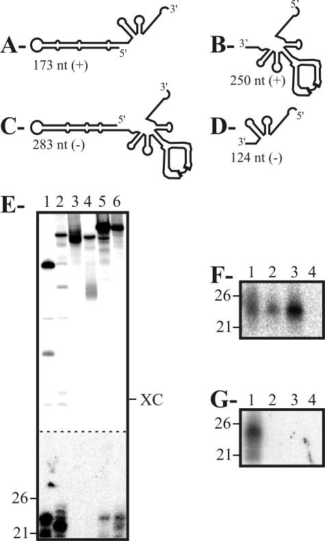
Dicer activity assays performed with various PLMVd-derived fragments. (A to D) Schematic representations of the plus-polarity (A, B) and minus-polarity (C, D) PLMVd fragments tested. The length, in nucleotides, is indicated for each fragment. (E) Autoradiogram of a PAGE gel of Dicer activity assays performed as described above. Lanes 1 to 6 are the 173-nt (lane 1), 283-nt (lane 2), 250-nt (lane 3), 124-nt (lane 4), and 338-nt fragments of plus and minus polarity (lanes 5 and 6, respectively). Adjacent to the gel, the positions of the RNA markers of 21 and 26 nt are indicated. XC indicates xylene cyanol. The bottom part (below the dashed line) has been overexposed in order to reveal the bands corresponding to the small products. (F, G) Autoradiograms of Northern blot hybridizations of cleavage assays performed in the presence of nonradioactive PLMVd RNA fragments of plus polarity. The conditions for hybridization have been described previously (12). The 5′-32P-oligonucleotides used as probes were complementary to a part of the P11 hairpin (5′-1GCCCACTGATGAGCCGCTGAAATGCGGCGAAACTTTTGAT40-3′) (F) and the right domain (5′-142CCGCTTGGTTCCCGAAGGAAAAGTCCCACCTTACCTCATTG182-3′) (G) of PLMVd. Lanes 1 to 4 are the duplexes of PLMVd strands of plus and minus polarity (lane 1), the 338-nt fragments (lane 2), and the 173-nt (lanes 3) and 250-nt (lane 4) fragments of plus polarity. Adjacent to the gel, the positions of the RNA markers of 21 and 26 nt are indicated.
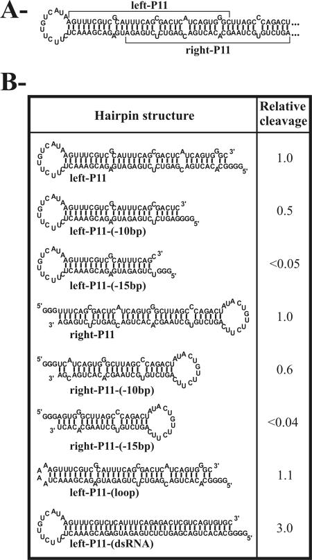
Dicer activity assays performed with various transcripts derived from the P11 hairpin. (A) The sequence and secondary structure of the hairpin is illustrated. This is a variant sequence of the pPD1 insert sequence (see reference 2). Three guanosines were added at the 5′ end of each transcript to ensure efficient transcription from the oligonucleotides. (B) The Dicer assays were performed as described in the text. The cleavage levels were determined and are reported as relative values in comparison to a value of 1.0 that was arbitrarily assigned to the left-P11 substrate.

Autoradiogram of a 10% PAGE gel of primer extension by the RdRP in wheat germ extract. Lane 1 is the extension of primer P11 (5′-GGG6UUUGAUGAUAUGAGUUUCGUC324-3′) on the 338-nt PLMVd template. Lane 2 is the extension of primer P10 (5′-GGG290ACUCAUCUUCCAGAAUCACU270-3′) on the 250-nt PLMVd fragments. Lanes 3 and 4 are controls with DNase digestion and without template, respectively. To the left of the gel, the positions of the RNA markers are indicated. XC indicates xylene cyanol.
References
Publication types
MeSH terms
Substances
LinkOut - more resources
Full Text Sources
