Comparative Study
. 2005 Apr 27;25(17):4232-42.
doi: 10.1523/JNEUROSCI.4957-04.2005.

Nerve growth factor rapidly increases muscarinic tone in mouse medial septum/diagonal band of Broca

Affiliations
Comparative Study

Nerve growth factor rapidly increases muscarinic tone in mouse medial septum/diagonal band of Broca

Chia-wen K Wu et al. J Neurosci. .

Abstract

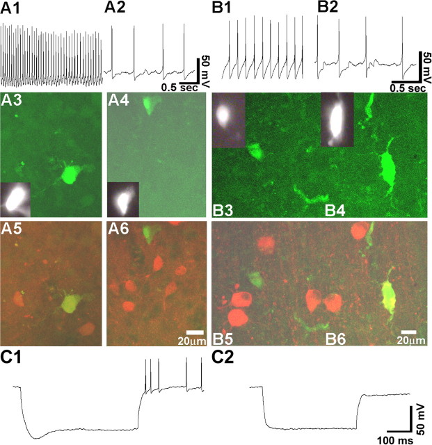
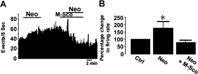
Nerve growth factor (NGF) has been implicated in maintaining and regulating normal functioning of the septohippocampal pathway. However, many aspects of its physiological actions and the underlying mechanisms await elucidation. In this study, we investigated the effect of acute NGF exposure on neurons in the mouse medial septum/diagonal band of Broca (MS/DB), focusing on the cholinergic neurons and the subpopulation of noncholinergic neurons that were identified to be putatively GABAergic. We report that MS/DB neurons in a thin slice preparation, when exposed to NGF via bath perfusion, rapidly and indiscriminately increased the rate of spontaneous firing in all MS/DB neurons. However, focal application of NGF to individual MS/DB neurons increased spontaneous firing in cholinergic, but not in the noncholinergic, subpopulation. The NGF-induced effect on cholinergic neurons was direct, requiring activation and signaling via TrkA receptors, which were immunohistochemically localized to the cholinergic neurons in the MS/DB. TrkA receptors were absent in putative GABAergic MS/DB neurons, and blockade of TrkA signaling in these and other noncholinergic neurons had no effect on their firing activity after exposure to NGF. Conversely, methyl scopolamine, blocked the increased firing activity of noncholinergic neurons during bath perfusion of NGF. We propose a cell type-specific mode of action for NGF in the MS/DB. The neurotrophin directly enhances cholinergic neuronal activity in the MS/DB through TrkA-mediated signaling, increasing acetylcholine release and, thus, muscarinic tone. This increase in muscarinic tone, in turn, results in heightened firing activity in noncholinergic MS/DB neurons.


Figures

Figure 1.
Figure 1.
Bath perfusion of NGF increases firing in MS/DB neurons. A, B, Representative rate-meter records of two MS/DB neurons displaying a slow (A) and fast (B) rate of spontaneous firing before [control (Ctrl)], during, and after bath perfusion of NGF (100 ng/ml) perfusion. C, Normalized histogram summarizing mean firing rate of all of the recorded neurons. The spontaneous firing rate was significantly faster when assessed at 3 and 8 min after NGF perfusion (*p < 0.05; Student's t test).
Figure 2.
Figure 2.
Fast-firing neurons are immunopositive for GABAergic markers, and slow-firing neurons are immunopositive for ChAT. Firing activity and parvalbumin/calbindin/calretinin (A) or ChAT (B) immunoreactivity in a fast-firing and slow-firing cell. A1, A2, Representative traces of spontaneous firing corresponding to a fast-firing and a slow-firing MS/DB neuron, respectively. A3, A4, Identification of the fast-firing cell and slow-firing cell with Lucifer yellow, respectively, first during recording (insets) and then after recovery in the fixed slice. A5, A6, The fixed slices were processed immunohistochemically using a mixture of parvalbumin/calbindin/calretinin antibodies. Overlay images show immunolabeling in the fast-firing cell (A5) but not in the slow-firing cell (A6). B1, B2, Representative traces of spontaneous firing corresponding to a fast-firing and a slow-firing cell, respectively. The two cells were recorded within the same microscopic field. B3, B4, The fast-firing cell, marked with Lucifer yellow during recording (B3, inset), is located on the left, and the slow-firing cell (B4, inset) is located on the right. Both neurons were recovered after fixation. B5, B6, The slow-firing cell (B6), but not the fast-firing cell (B5), was ChAT immunoreactive. C1, C2, Responses of a noncholinergic and a cholinergic neuron, respectively, to a hyperpolarizing current step. Note the depolarization sag in the noncholinergic neuron. Calibration applies to both C1 and C2.
Figure 3.
Figure 3.
Focal application of NGF acutely increases firing in cholinergic, but not noncholinergic, neurons. A, B, Representative rate-meter records of slow-firing and fast-firing neurons, respectively, illustrate spontaneous firing before, during, and after NGF (100 ng/ml) application. Insets show digitized traces from the same cells. Calibration: 0.5 s (A) and 1 s (B), 50 mV. Ctrl, Control. C, D, Summary graph of the effect of NGF on cholinergic and noncholinergic neurons, respectively. Each symbol represents data obtained from one neuron. The 45° equivalence line represents the result expected if the firing rate remains unchanged after application of NGF. The shaded area represents a 10% increase or decrease and reflects the range of variability normally encountered while monitoring the rate of spontaneous firing in MS/DB neurons. Most of the slow-firing neurons show increased firing rates, whereas fast-firing neurons showed more variable responses. E, F, Normalized histograms summarizing mean firing rates of cholinergic (n = 15) and noncholinergic (n = 11) neurons in the presence of 10 or 100 ng/ml NGF. During acute and focal NGF exposure, the mean rate of spontaneous firing in cholinergic neurons was significantly faster in both concentrations (p < 0.05; Student's t test) than that during the control (pre-NGF exposure) period, whereas that of noncholinergic neurons was unaffected (p > 0.05; Student's t test). Asterisks in E denote statistical significance.
Figure 4.
Figure 4.
MS/DB neurons do not coexpress TrkA and GABAergic markers. Confocal images taken from histological sections containing MS/DB double labeled using a mixture of antibodies against parvalbumin and calbindin (A) and TrkA receptor (B). C, Overlay of A and B. Representative fields in A1, B1, and C1 are shown at higher magnification in A2, B2, and C2, respectively.
Figure 5.
Figure 5.
Cholinergic neurons, but not noncholinergic neurons, respond to acute NGF application and display TrkA immunoreactivity. A, E, Two separate cells filled with Lucifer yellow during recording (insets) and recovered after fixation of the slices. B, F, Only the cell shown in B is immunoreactive to the antibody against TrkA. C, G, Overlay images of A, B and E, F, respectively. D, H, Representative traces of the cholinergic and noncholinergic neuron shown in A and E, respectively, of spontaneous firing before [control (Ctrl)], during, and after [recovery (Rec)] NGF application.
Figure 6.
Figure 6.
K252a inhibits NGF-induced increase in firing of cholinergic neurons. A, Rate-meter record from a cholinergic neuron illustrates that K252a (200 nm) prevented the increase in firing normally observed during application of NGF (100 ng/ml), which, in the same cell, persisted in the presence of K252b (200 nm). B, Normalized histograms summarizing that K252a, but not K252b, blocks the NGF-induced increase in mean firing rates of cholinergic neurons. During exposure to K252a, the NGF-induced mean firing rate of cholinergic neurons was not significantly different from that monitored before NGF application when slices were exposed only to K252 (p > 0.05; Student's t test), whereas the NGF-induced change in mean firing rate of cholinergic neurons was statistically significant in the presence of K252b (p < 0.05; Student's t test). C, Phosphorylated TrkA was detected in tissue microdissected from the MS/DB after NGF perfusion. D, Normalized histogram summarizing mean density of Western blots obtained from MS/DB tissues. MS/DB tissues perfused with NGF displayed significantly stronger signal than that perfused with aCSF (p < 0.05; Student's t test; n = 5). Asterisks in B and D denote statistical significance. Ctrl, Control tissue processed in parallel without incubation with NGF.
Figure 7.
Figure 7.
TrkA-Fc inhibits the acute effect of NGF on firing of cholinergic neurons. A, C, Representative rate-meter records of cholinergic neurons demonstrate spontaneous firing before, during, and after focal NGF (100 ng/ml) application in the presence of TrkA-Fc and boiled TrkA-Fc, respectively. The cell in A also showed increased firing in the presence of NGF only. B, D, Normalized histograms summarizing that TrkA-Fc, but not boiled TrkA-Fc, blocks the NGF-induced increase in mean firing rates of cholinergic neurons. In the presence of TrkA-Fc, the NGF-induced increase in mean firing rate of cholinergic neurons was not significantly different from that monitored during the TrkA-Fc only period (p > 0.05; n = 7). The NGF effect resumed during washout of TrkA-Fc (p < 0.05). Boiled TrkA-Fc did not affect NGF-induced increase firing (p < 0.05; Student's t test; n = 6). Asterisks in B and D denote statistical significance.
Figure 8.
Figure 8.
The NGF-induced effect on firing of cholinergic MS/DB neurons persists in p75NTR-/- mice. A, Representative rate-meter record of a cholinergic neuron from a p75NTR-/- mouse demonstrates spontaneous firing before, during, and after focal NGF (100 ng/ml) application. B, Summary histogram indicates that, under p75NTR-/- conditions, NGF application significantly increased firing rate of MS/DB cholinergic neurons by 94 ± 45% (n = 12; mean ± SEM; p < 0.05; Student's t test). Asterisk denotes statistical significance. Ctrl, Control period before NGF application.
Figure 9.
Figure 9.
Neostigmine increases ambient acetylcholine in mouse MS/DB. A, C, Representative rate-meter records (top) and digitized traces (bottom) from a noncholinergic (A) and cholinergic (C) neuron, illustrating spontaneous firing rates before [control (Ctrl)], during, and after [recovery (Rec)] neostigmine (Neo) application. B, D, Normalized histograms summarizing mean spontaneous firing rates of noncholinergic (B) and cholinergic (D) neurons in the presence of neostigmine. The mean firing rate of noncholinergic neurons during exposure to neostigmine was significantly faster than that during the control period (n = 16; mean ± SEM; p < 0.05; Student's t test), whereas that of cholinergic neurons was unaffected (n = 15; mean ± SEM; p > 0.05; Student's t test). The asterisk in B denotes statistical significance.
Figure 10.
Figure 10.
Methyl scopolamine blocks the neostigmine-induced increase in firing in noncholinergic neurons. A, A representative rate-meter record displaying spontaneous firing of a noncholinergic neuron before [control (Ctrl)], during, and after neostigmine (Neo) application either alone or in conjunction with methyl scopolamine (M-Sco). B, Histogram summarizing the normalized mean firing rate of noncholinergic neurons (n = 6) during neostigmine exposure without and with concomitant application of methyl scopolamine. With neostigmine alone, the mean firing rate of noncholinergic neurons was significantly faster (78.2 ± 45%; mean ± SEM; p < 0.05; Student's t test). The neostigmine-induced increase in firing was blocked in the presence of methyl scopolamine. The asterisk in B denotes statistical significance.
Figure 11.
Figure 11.
Methyl scopolamine blocks the increased firing in noncholinergic but not cholinergic neurons in response to bath perfusion of NGF. A, B, Representative rate-meter records of a noncholinergic and a cholinergic neuron, respectively, display spontaneous firing before (Ctrl, control), during, and after whole-slice NGF (100 ng/ml) perfusion in the presence of methyl scopolamine (M-Sco; 10 μm). C, Normalized histograms summarizing mean firing rates of noncholinergic, putatively GABAergic (n = 8), and cholinergic (n = 6) neurons after NGF perfusion in the presence of methyl scopolamine. The mean rate of spontaneous firing in cholinergic neurons was significantly faster (p < 0.05; Student's t test) than that during the control (pre-NGF/methyl scopolamine exposure) period, whereas that of noncholinergic neurons was abolished by methyl scopolamine (p > 0.05; Student's t test). The asterisk in B denotes statistical significance.

Similar articles

Cited by

References

    1. Acsády L, Pascual M, Rocamora N, Soriano E, Freund TF (2000) Nerve growth factor but not neurotrophin-3 is synthesized by hippocampal GABAergic neurons that project to the medial septum. Neuroscience 98: 23-31. - PubMed
    1. Alreja M, Wu M, Liu W, Atkins JB, Leranth C, Shanabrough M (2000) Muscarinic tone sustains impulse flow in the septohippocampal GABA but not cholinergic pathway: implication for learning and memory. J Neurosci 20: 8103-8110. - PMC - PubMed
    1. Andersen P, Bland HB, Myhrer T, Schwartzkroin PA (1979) Septohippocampal pathway necessary for dentate theta production. Brain Res 165: 13-22. - PubMed
    1. Backman C, Rose GM, Hoffer BJ, Henry MA, Bartus RT, Friden P, Granholm AC (1996) Systemic administration of a nerve growth factor conjugate reverses age-related cognitive dysfunction and prevents cholinergic neuron atrophy. J Neurosci 16: 5437-5442. - PMC - PubMed
    1. Bamji SX, Majdan M, Pozniak CD, Belliveau DJ, Aloyz R, Kohn J, Causing CG, Miller FD (1998) The p75 neurotrophin receptor mediates neuronal apoptosis and is essential for naturally occurring sympathetic neuron death. J Cell Biol 140: 911-923. - PMC - PubMed

Publication types

MeSH terms

LinkOut - more resources