Comparative Study
. 2005 Apr 27;25(17):4353-64.
doi: 10.1523/JNEUROSCI.0885-05.2005.

Aldolases a and C are ribonucleolytic components of a neuronal complex that regulates the stability of the light-neurofilament mRNA

Affiliations
Comparative Study

Aldolases a and C are ribonucleolytic components of a neuronal complex that regulates the stability of the light-neurofilament mRNA

Rafaela Cañete-Soler et al. J Neurosci. .

Erratum in

  • J Neurosci. 2006 May 10;26(19):5276

Abstract

A 68 nucleotide segment of the light neurofilament (NF-L) mRNA, spanning the translation termination signal, participates in regulating the stability of the transcript in vivo. Aldolases A and C, but not B, interact specifically with this segment of the transcript in vitro. Aldolases A and C are glycolytic enzymes expressed in neural cells, and their mRNA binding activity represents a novel function of these isozymes. This unsuspected new activity was first uncovered by Northwestern blotting of a brainstem/spinal cord cDNA library. It was confirmed by two-dimensional fractionation of mouse brain cytosol followed by Northwestern hybridization and protein sequencing. Both neuronal aldolases interact specifically with the NF-L but not the heavy neurofilament mRNA, and their binding to the transcript excludes the poly(A)-binding protein (PABP) from the complex. Constitutive ectopic expression of aldolases A and C accelerates the decay of a neurofilament transgene (NF-L) driven by a tetracycline inducible system. In contrast, mutant transgenes lacking mRNA sequence for aldolase binding are stabilized. Our findings strongly suggest that aldolases A and C are regulatory components of a light neurofilament mRNA complex that modulates the stability of NF-L mRNA. This modulation likely involves endonucleolytic cleavage and a competing interaction with the PABP. Interactions of aldolases A and C in NF-L expression may be linked to regulatory pathways that maintain the highly asymmetrical form and function of large neurons.


Figures
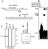
Figure 1.
Figure 1.
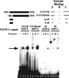
Detection of specific interactions between a 68 nt segment of neurofilament RNA and cytoplasmic extract from mouse tissues. A, Schematic diagram of NF-L mRNA showing location and sequence of destabilizing element. B, Electrophoretic mobility shift assay after incubation at 37°C of 1 ng of radiolabeled NF-L 68 with 50 μg of brain (lane 2) or liver (lane 3) extracts for 5 min before digestion with RNase T1. Binding reactions were analyzed on a native low-ionic strength 5% polyacrylamide gel. C, Identification of polypeptides in a brain extract RNA-protein complex by Northwestern analysis. A binding reaction identical to that shown in B (lane 2) was performed with unlabeled specific RNA probe and brain extract and resolved on a native 5% gel. The complex was excised from the gel, and the proteins were eluted and fractionated in a 10% SDS-PAGE. D, Western blot analysis of brain extract RNA-protein complex. Proteins from brain RNA-protein complex in B (lane 2) were immunoblotted (IB) using anti-aldolase antibody. A control normal rabbit serum (NRS) is shown in lane 1, protein from brain ribonucleoprotein complex is shown in lane 2, and control purified aldolase is shown in lane 3.
Figure 2.
Figure 2.
Identification of neuronal aldolases A and C as components of a brain extract neurofilament RNA-protein complex. A, 2D-PAGE and Northwestern analysis of proteins from 50 μg of mouse brain cytoplasmic extract stained with silver (top) or transferred to nitrocellulose and hybridized separately with an NF-L 68 nt RNA probe (middle) or a nonspecific SK + 70 (bottom) identified two 39 kDa RNA-binding proteins (arrowhead within the gel). The left lane in the middle panel shows hybridization with two 39 kDa proteins in mouse brain proteins separated by 1D electrophoresis. The polypeptides present in the two spots were digested within gel slices with trypsin and extracted. B, HPLC chromatograms of gel-eluted peptides in A from the right spot (R) or the left spot (L). C, MALDI of HPLC fractions selected for Edman sequencing. Sequences (listed above) matched those of aldolase C (fractions 36 and 42) and aldolase A (fraction 66). m/z, Mass-to-charge ratio.
Figure 3.
Figure 3.
Isozyme-dependent formation of neurofilament RNA-protein complexes. Binding reactions were performed as described in Figure 1. A, Electrophoretic mobility shift assay of internally radiolabeled NF-68 RNA with 0-80 μg of brain extract (lanes 1-4) or with 60-200 nm purified aldolase C (lanes 5-8). B, Formation of isozyme-dependent complexes after incubation of radiolabeled NF-L68 RNA with 10 nm purified aldolase A (lane 2), aldolase B (lane 3), or aldolase C (lane 4). C, Isozyme dose-response and magnesium requirements for complex formation. A total of 10-80 nm purified aldolase A (lanes 2-5), aldolase B (lanes 6-9), and aldolase C (lanes 10-13) was incubated with capped internally labeled NF-68 RNA, and complexes were resolved in a nondenaturing 5% acrylamide gel. Parallel binding reactions in the presence of 30 mm EDTA were conducted with 20-80 nm aldolase A (lanes 14-16) or aldolase C (lanes 17-19).
Figure 4.
Figure 4.
Specificity of formation of aldolase-neurofilament RNA complexes by electrophoretic mobility shift assays and circular dichroism spectroscopy. A, Electrophoretic mobility shift assay after incubation of uncapped radiolabeled NF-L68 with aldolase C in the presence of 0- to 100-fold excess of unlabeled NF-L68 (lanes 3-7), 0- to 800-fold of unrelated SV40 (lanes 8-13), or 40- to 200-fold of TNFα-ARE (lanes 14-16) or IL-3 ARE RNAs (lanes 17-19). The arrow denotes shifted complexes. B, C, Near-UV CD spectra from 240-320 nm for RNAs alone, nonspecific SK + 70 RNA (B) or with specific NF-L 68 RNA (C), or with aldolase A (Aldol A). D, Far-UV CD spectra of aldolases A (Aldol A) and C (Aldol C) alone or in complex with NF-L 68 RNA. The CD intensity was monitored as a function of temperature at 222 nm.
Figure 5.
Figure 5.
Mapping of protein-binding in the 680 nt mouse neurofilament light stability determinant region. The 680 nt encompasses the full 3′-UTR plus 250 nt of distal coding region of the neurofilament light subunit sequence. Binding reactions were conducted as described in Figure 1. Electrophoretic mobility shift assays of radiolabeled 0.68 kb RNA sequence (lane 1) or shorter variants depicted in the top panel (lanes 5, 9, 13, 17, 21, 25) were conducted to test for their ability to form band-shifted complexes with 150 ng of purified aldolase A (lanes 2, 6, 10, 14, 18, 22, 26), aldolase B (lanes 3, 7, 11, 15, 19, 23, 27), or aldolase C (lanes 4, 8, 12, 16, 20, 24, 28). Arrows denote the position of the band-shifted complexes.
Figure 6.
Figure 6.
Aldolase-isozyme binding to the 3′-UTR of the human NF-L (hNF-L) (mRNA). Electrophoretic mobility shift assays of radiolabeled human NF-L 3′-UTR (lanes 1-4) or a variant lacking 45 nt of proximal 3′-UTR (lanes 5-8) or a 45 nt from mouse (lanes 9-12) or human proximal 3′-UTR (lanes 13-16) depicted in the top panel were conducted to test for their ability to form band-shifted complexes with 150 ng of purified aldolase A (lanes 2, 6, 10, 14), aldolase B (lanes 3, 7, 11, 15), or aldolase C (lanes 4, 8, 12, 16). Arrows denote the position of the band-shifted complexes. mNF-L, Mouse NF-L.
Figure 7.
Figure 7.
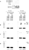
Detection in vivo of direct specific interactions between the NF-L mRNA and aldolases A and C in Neuro-2a cells. A, Western blot analyses after HA immunoprecipitation of ribonucleoprotein complexes from cells expressing NF-L and HA-tagged aldolases A and C. IB, Immunoblot; Ip, immunopellet; Ipp, immunoprecipitation; L, lysate. B-D, RT-PCR amplification of RNA extracted from immunoprecipitates (lanes 1-3 and 7-9) or one-tenth of total RNP lysate (lanes 4-6 and 10-12) of cells transfected with vector alone (lanes 1, 4, 7, 10) or cotransfected with NF-L and HA-aldolase A (lanes 2, 5, 8, 11) or HA-aldolase C (lanes 3, 6, 9, 12). Primers for cDNA amplification were from transgene NF-L and vector (B), endogenous NF-H (C), and v-erb (D) sequences. All reactions contained 0.1 μm respective end-labeled sense primer. Sample aliquots were withdrawn after 12 and 22 cycles and fractionated in polyacrylamide gels along with radioactive DNA markers.
Figure 8.
Figure 8.
mRNA decay assay of endogenous NF mRNAs or inducible NF-L transgene in the presence of ectopic aldolase in Neuro-2a cells. A, Ribonuclease protection assay of NF- and β-actin (β-A) mRNA levels. Target NF-L transgene was activated for 3 h in the absence of doxycycline and then inactivated by the addition of 0.5 μg/ml ligand (see Materials and Methods). B, Quantitation of NF levels normalized to levels of β-actin mRNA. Data represent the mean of three consecutive independent transfections. H, NF-H; L, NF-L; M, NF-M.
Figure 9.
Figure 9.
Effect of constitutive ectopic expression of aldolases A and C on the decay of inducible NF-L mRNA and mutant transgenes in COS cells. A, Ribonuclease protection assay of NF-L and β-actin mRNA levels. Target neurofilament transgenes were activated for 3 h in the absence of doxycycline and then inactivated by the addition of 0.5 μg/ml ligand (see Materials and Methods). B, Quantitation of NF-L mRNA levels normalized to levels of β-actin mRNA. Data represent the mean of three consecutive independent transfections. An, Poly(A) tail; CRD, coding region deleted; UTRD, UTR deleted.
Figure 10.
Figure 10.
Analysis of NF mRNA protein:protein interactions in cells coexpressing the NF-L and aldolases. A, Anti-PABP coimmunoprecipitation assay of RNP complexes from COS cells transfected with indicated plasmids. The immunoprecipitation (Ip) was performed with a monoclonal anti-PABP and immunoblotted (IB) with polyclonals anti-PABP or anti-aldolase C. Specific immunodetection of endogenous PABP (lanes 1-8) and aldolase shows a competing interaction between the two factors. B, Anti-HA coimmunoprecipitation assay of RNPs from COS cells coexpressing the same constructs as in A. L, Lysate.
Figure 11.
Figure 11.
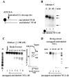
Ribonucleolytic activity of aldolase. A, Diagram of NF-L transcripts used in degradation assays. NF-L 68 encompasses 23 nt of distal coding region and 45 nt of proximal 3′-UTR, cap-labeled with vaccinia-guanyltransferase and P32 α-GTP (top diagram) or uncapped but end-labeled with P32 from γ-ATP (bottom diagram). The arrow indicates a potential cleavage site. B, Degradation assay after incubation of 100 nm purified aldolase C with cap-labeled NF-L68. C, Degradation assay after incubation of uncapped end-labeled NF-68 with 100 nm aldolase C in the absence (lane 3) or presence of increasing amounts of brain extract (lanes 4-8). Quantitation of input uncapped NF-68 and intermediates as a function of added brain extract concentration (see Results). D, Degradation assay of uncapped end-labeled NF-L 68 by activities present in brain extract.

Similar articles

Cited by

References

    1. Ahn AH, Dziennis S, Hawkes R, Herrup K (1994) The cloning of zebrin II reveals its identity with aldolase C. Development 120: 2081-2090. - PubMed
    1. Amrani N, Ganesan R, Kervestin S, Mangus DA, Ghosh S, Jacobson A (2004) A faux 3′-UTR promotes aberrant termination and triggers non-sense-mediated mRNA decay. Nature 432: 118-129. - PubMed
    1. Antic D, Lu N, Keene JD (1999) ELAV tumor antigen, Hel-N1, increases translation of neurofilament M mRNA and induces formation of neurites in human teratocarcinoma cells. Genes Dev 13: 449-461. - PMC - PubMed
    1. Baraniak AP, Lasda EL, Wagner EJ, García-Blanco M (2003) A stem structure in fibroblast growth factor receptor 2 transcripts mediates cell type-specific splicing by approximating intronic control elements. Mol Cell Biol 23: 9327-9337. - PMC - PubMed
    1. Berardini TZ, Drygas-Williams M, Callard GV, Tolan DR (1997) Identification of neuronal isozyme specific residues by comparison of goldfish aldolase C to other aldolases. Comp Biochem Physiol 117A: 471-476. - PubMed

Publication types

MeSH terms

LinkOut - more resources