

Processing of radical prostatectomy specimens for correlation of data from histopathological, molecular biological, and radiological studies: a new whole organ technique
- PMID: 15858122
- PMCID: PMC1770644
- DOI: 10.1136/jcp.2004.021808
Processing of radical prostatectomy specimens for correlation of data from histopathological, molecular biological, and radiological studies: a new whole organ technique
Abstract
Aims: To develop a method of processing non-formalin fixed prostate specimens removed at radical prostatectomy to obtain fresh tissue for research and for correlating diagnostic and molecular results with preoperative imaging.
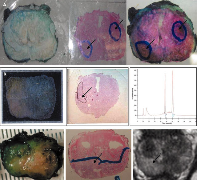
Methods/results: The method involves a prostate slicing apparatus comprising a tissue slicer with a series of juxtaposed planar stainless steel blades linked to a support, and a cradle adapted to grip the tissue sample and receive the blades. The fresh prostate gland is held in the cradle and the blades are moved through the cradle slits to produce multiple 4 mm slices of the gland in a plane perpendicular to its posterior surface. One of the resulting slices is preserved in RNAlater. The areas comprising tumour and normal glands within this preserved slice can be identified by matching it to the haematoxylin and eosin stained sections of the adjacent slices that are formalin fixed and paraffin wax embedded. Intact RNA can be extracted from the identified tumour and normal glands within the RNAlater preserved slice. Preoperative imaging studies are acquired with the angulation of axial images chosen to be similar to the slicing axis, such that stained sections from the formalin fixed, paraffin wax embedded slices match their counterparts on imaging.
Conclusions: A novel method of sampling fresh prostate removed at radical prostatectomy that allows tissue samples to be used both for diagnosis and molecular analysis is described. This method also allows the integration of preoperative imaging data with histopathological and molecular data obtained from the prostate tissue slices.
Figures


Similar articles
-
Technical Note: Method to correlate whole-specimen histopathology of radical prostatectomy with diagnostic MR imaging.Med Phys. 2016 Mar;43(3):1065-72. doi: 10.1118/1.4941016. Med Phys. 2016. PMID: 26936694 Free PMC article.
-
Methods of radical prostatectomy specimen processing: a novel technique for harvesting fresh prostate cancer tissue and review of processing techniques.Mod Pathol. 1993 Mar;6(2):201-7. Mod Pathol. 1993. PMID: 8483892
-
Ceramic foam plates: a new tool for processing fresh radical prostatectomy specimens.Virchows Arch. 2014 Dec;465(6):637-42. doi: 10.1007/s00428-014-1665-8. Epub 2014 Oct 17. Virchows Arch. 2014. PMID: 25323812
-
Optimum slicing of radical prostatectomy specimens for correlation between histopathology and medical images.Int J Comput Assist Radiol Surg. 2010 Sep;5(5):471-87. doi: 10.1007/s11548-010-0405-z. Epub 2010 Feb 24. Int J Comput Assist Radiol Surg. 2010. PMID: 20180036 Review.
-
Handling of radical prostatectomy specimens.Histopathology. 2012 Jan;60(1):118-24. doi: 10.1111/j.1365-2559.2011.04002.x. Histopathology. 2012. PMID: 22212081 Review.
Cited by
-
A decade in prostate cancer: from NMR to metabolomics.Nat Rev Urol. 2011 May 17;8(6):301-11. doi: 10.1038/nrurol.2011.53. Nat Rev Urol. 2011. PMID: 21587223 Review.
-
Detection of TMPRSS2-ERG translocations in human prostate cancer by expression profiling using GeneChip Human Exon 1.0 ST arrays.J Mol Diagn. 2008 Jan;10(1):50-7. doi: 10.2353/jmoldx.2008.070085. Epub 2007 Dec 28. J Mol Diagn. 2008. PMID: 18165275 Free PMC article.
-
PEOPLE: PatiEnt prOstate samPLes for rEsearch, a tissue collection pathway utilizing magnetic resonance imaging data to target tumor and benign tissue in fresh radical prostatectomy specimens.Prostate. 2019 May;79(7):768-777. doi: 10.1002/pros.23782. Epub 2019 Feb 26. Prostate. 2019. PMID: 30807665 Free PMC article.
-
Liver-specific 3D sectioning molds for correlating in vivo CT and MRI with tumor histopathology in woodchucks (Marmota monax).PLoS One. 2020 Mar 26;15(3):e0230794. doi: 10.1371/journal.pone.0230794. eCollection 2020. PLoS One. 2020. PMID: 32214365 Free PMC article.
-
Customized Tool for the Validation of Optical Coherence Tomography in Differentiation of Prostate Cancer.Technol Cancer Res Treat. 2017 Feb;16(1):57-65. doi: 10.1177/1533034615626614. Epub 2016 Jul 7. Technol Cancer Res Treat. 2017. PMID: 26818025 Free PMC article.
References
-
- Cancer Research UK. Cancer Stats Incidence—UK, Vol. 6. London: Cancer Research UK, 2003.
-
- Parker C . Active surveillance: towards a new paradigm in the management of early prostate cancer. Lancet Oncol 2004;5:101–6. - PubMed
-
- Bagnall S, Klotz L. Conservative versus radical therapy of prostate cancer: how have recent advances in molecular markers and imaging enhanced our ability to prognosticate risk? Semin Oncol 2003;30:587–95. - PubMed
-
- Aihara M, Wheeler TM, Ohori M, et al. Heterogeneity of prostate cancer in radical prostatectomy specimens. Urology 1994;43:60–6. - PubMed
-
- Sakr WA, Grignon DJ. Prostate. Practice parameters, pathologic staging, and handling radical prostatectomy specimens. Urol Clin North Am 1999;26:453–63. - PubMed
Publication types
MeSH terms
Substances
LinkOut - more resources
Full Text Sources