

Clathrin is required for the function of the mitotic spindle
- PMID: 15858577
- PMCID: PMC3492753
- DOI: 10.1038/nature03502
Clathrin is required for the function of the mitotic spindle
Abstract
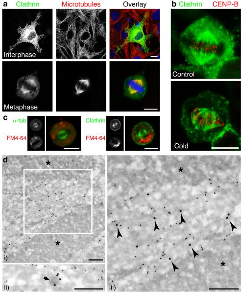
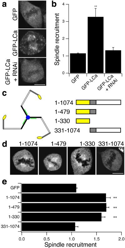
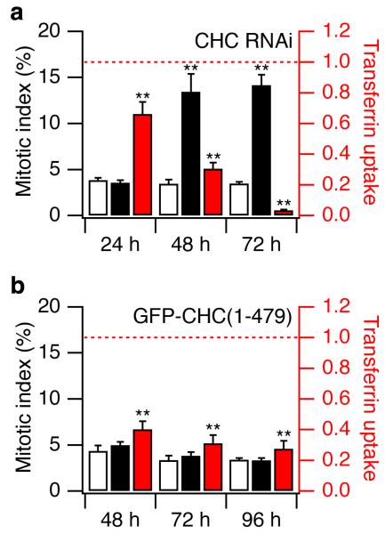
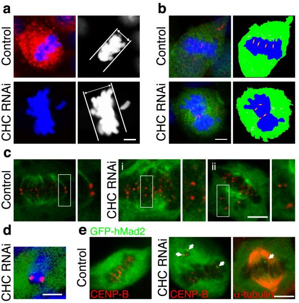
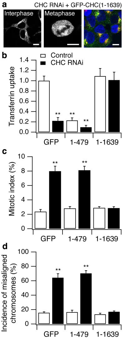
Clathrin has an established function in the generation of vesicles that transfer membrane and proteins around the cell. The formation of clathrin-coated vesicles occurs continuously in non-dividing cells, but is shut down during mitosis, when clathrin concentrates at the spindle apparatus. Here, we show that clathrin stabilizes fibres of the mitotic spindle to aid congression of chromosomes. Clathrin bound to the spindle directly by the amino-terminal domain of clathrin heavy chain. Depletion of clathrin heavy chain using RNA interference prolonged mitosis; kinetochore fibres were destabilized, leading to defective congression of chromosomes to the metaphase plate and persistent activation of the spindle checkpoint. Normal mitosis was rescued by clathrin triskelia but not the N-terminal domain of clathrin heavy chain, indicating that stabilization of kinetochore fibres was dependent on the unique structure of clathrin. The importance of clathrin for normal mitosis may be relevant to understanding human cancers that involve gene fusions of clathrin heavy chain.
Figures
Similar articles
-
Sorting nexin 9 recruits clathrin heavy chain to the mitotic spindle for chromosome alignment and segregation.PLoS One. 2013 Jul 5;8(7):e68387. doi: 10.1371/journal.pone.0068387. Print 2013. PLoS One. 2013. PMID: 23861900 Free PMC article.
-
Trimerisation is important for the function of clathrin at the mitotic spindle.J Cell Sci. 2006 Oct 1;119(Pt 19):4071-8. doi: 10.1242/jcs.03192. Epub 2006 Sep 12. J Cell Sci. 2006. PMID: 16968737 Free PMC article.
-
Clathrin's adaptor interaction sites are repurposed to stabilize microtubules during mitosis.J Cell Biol. 2020 Feb 3;219(2):e201907083. doi: 10.1083/jcb.201907083. J Cell Biol. 2020. PMID: 31932847 Free PMC article.
-
The role of clathrin in mitotic spindle organisation.J Cell Sci. 2012 Jan 1;125(Pt 1):19-28. doi: 10.1242/jcs.094607. J Cell Sci. 2012. PMID: 22294613 Free PMC article. Review.
-
Structural insights into the clathrin coat.Semin Cell Dev Biol. 2007 Aug;18(4):448-58. doi: 10.1016/j.semcdb.2007.07.006. Epub 2007 Aug 16. Semin Cell Dev Biol. 2007. PMID: 17702618 Review.
Cited by
-
A Differential Protein Study on Bronchoalveolar Lavage Fluid at Different Stages of Silicosis.Comb Chem High Throughput Screen. 2024;27(16):2366-2401. doi: 10.2174/0113862073260760231023055036. Comb Chem High Throughput Screen. 2024. PMID: 38173059
-
Oocyte spindle proteomics analysis leading to rescue of chromosome congression defects in cloned embryos.J Proteome Res. 2010 Nov 5;9(11):6025-6032. doi: 10.1021/pr100827j. Epub 2010 Oct 20. J Proteome Res. 2010. PMID: 20883044 Free PMC article.
-
The Cdc42 GEF Intersectin 2 controls mitotic spindle orientation to form the lumen during epithelial morphogenesis.J Cell Biol. 2010 May 17;189(4):725-38. doi: 10.1083/jcb.201002047. J Cell Biol. 2010. PMID: 20479469 Free PMC article.
-
The endocytic matrix.Nature. 2010 Jan 28;463(7280):464-73. doi: 10.1038/nature08910. Nature. 2010. PMID: 20110990 Review.
-
Linked in: formation and regulation of microtubule attachments during chromosome segregation.Curr Opin Cell Biol. 2014 Feb;26:113-22. doi: 10.1016/j.ceb.2013.12.005. Epub 2014 Jan 7. Curr Opin Cell Biol. 2014. PMID: 24529253 Free PMC article. Review.
References
-
- Kirchhausen T. Clathrin. Annu. Rev. Biochem. 2000;69:699–727. - PubMed
-
- Brodsky FM, Chen CY, Knuehl C, Towler MC, Wakeham DE. Biological basket weaving: formation and function of clathrin-coated vesicles. Annu. Rev. Cell Dev. Biol. 2001;17:517–68. - PubMed
-
- Robinson MS. Adaptable adaptors for coated vesicles. Trends Cell Biol. 2004;14:167–74. - PubMed
-
- Fotin A, et al. Molecular model for a complete clathrin lattice from electron cryomicroscopy. Nature. 2004;432:573–9. - PubMed
-
- Gaidarov I, Santini F, Warren RA, Keen JH. Spatial control of coated-pit dynamics in living cells. Nat. Cell Biol. 1999;1:1–7. - PubMed
Publication types
MeSH terms
Substances
Grants and funding
LinkOut - more resources
Full Text Sources
Other Literature Sources
Molecular Biology Databases
