Representations of faces and body parts in macaque temporal cortex: a functional MRI study
- PMID: 15860578
- PMCID: PMC1100800
- DOI: 10.1073/pnas.0502605102
Representations of faces and body parts in macaque temporal cortex: a functional MRI study
Abstract
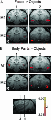
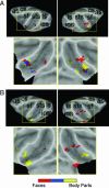
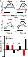
Human neuroimaging studies suggest that areas in temporal cortex respond preferentially to certain biologically relevant stimulus categories such as faces and bodies. Single-cell studies in monkeys have reported cells in inferior temporal cortex that respond selectively to faces, hands, and bodies but provide little evidence of large clusters of category-specific cells that would form "areas." We probed the category selectivity of macaque temporal cortex for representations of monkey faces and monkey body parts relative to man-made objects using functional MRI in animals trained to fixate. Two face-selective areas were activated bilaterally in the posterior and anterior superior temporal sulcus exhibiting different degrees of category selectivity. The posterior face area was more extensively activated in the right hemisphere than in the left hemisphere. Immediately adjacent to the face areas, regions were activated bilaterally responding preferentially to body parts. Our findings suggest a category-selective organization for faces and body parts in macaque temporal cortex.
Figures


References
-
- Haxby, J. V., Gobbini, M. I., Furey, M. L., Ishai, A., Schouten, J. L. & Pietrini, P. (2001) Science 293, 2425–2430. - PubMed
-
- Grill-Spector, K. & Malach, R. (2004) Annu. Rev. Neurosci. 27, 649–677. - PubMed
-
- McCarthy, G., Puce, A., Gore, J. C. & Allison, T. (1997) J. Cognit. Neurosci. 9, 605–610. - PubMed
Publication types
MeSH terms
Grants and funding
LinkOut - more resources
Full Text Sources
Other Literature Sources
Medical

