. 2005 May 10;102(19):6771-6.
doi: 10.1073/pnas.0408135102. Epub 2005 Apr 28.

Interfacial folding and membrane insertion of designed peptides studied by molecular dynamics simulations

Affiliations

Interfacial folding and membrane insertion of designed peptides studied by molecular dynamics simulations

Wonpil Im et al. Proc Natl Acad Sci U S A. .

Abstract

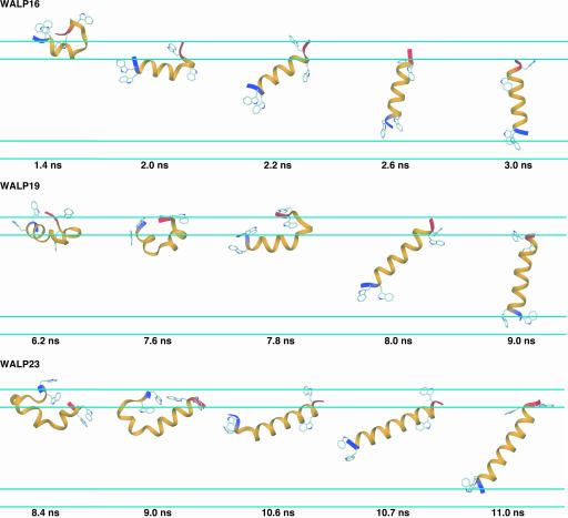
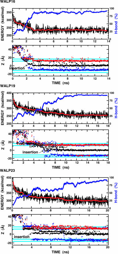
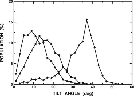
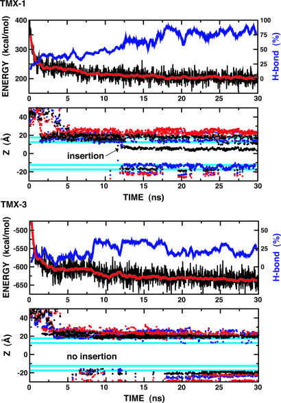
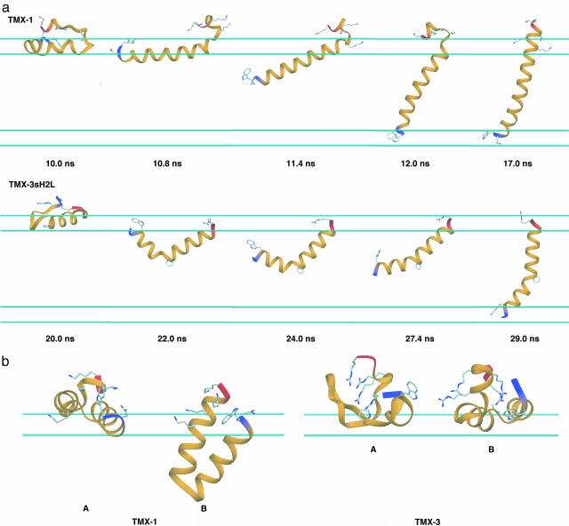
The mechanism of interfacial folding and membrane insertion of designed peptides is explored by using an implicit membrane generalized Born model and replica-exchange molecular dynamics. Folding/insertion simulations initiated from fully extended peptide conformations in the aqueous phase, at least 28 A away from the membrane interface, demonstrate a general mechanism for structure formation and insertion (when it occurs). The predominately hydrophobic peptides from the synthetic WALP and TMX series first become localized at the membrane-solvent interface where they form significant helical secondary structure via a helix-turn-helix motif that inserts the central hydrophobic residues into the membrane interior, and then fluctuations occur that provide a persistent helical structure throughout the peptide and it inserts with its N-terminal end moving across the membrane. More specifically, we observed that: (i) the WALP peptides (WALP16, WALP19, and WALP23) spontaneously insert in the membrane as just noted; (ii) TMX-1 also inserts spontaneously after a similar mechanism and forms a transmembrane helix with a population of approximately 50% at 300 K; and (iii) TMX-3 does not insert, but exists in a fluctuating membrane interface-bound form. These findings are in excellent agreement with available experimental data and demonstrate the potential for new implicit solvent/membrane models together with advanced simulation protocols to guide experimental programs in exploring the nature and mechanism of membrane-associated folding and insertion of biologically important peptides.


Figures

Fig. 1.
Fig. 1.
Characteristic conformations for one replica of each of the WALP peptides illustrate the conformational and configurational changes that occur during membrane insertion. The N terminus is blue, and the C terminus is red. Trp residues are shown in ball-and-stick representations, and the cyan lines represent the 5-Å membrane smoothing regions.
Fig. 2.
Fig. 2.
Time series for several properties for each WALP peptide from the lowest temperature (300 K) ensemble during the REX simulations. (Upper) Profiles of total potential energy (black) as a function of time and its running average over 400-ps windows. The blue lines represent a running average of backbone H-bond fractions, defined as the number of H bonds in each configuration divided by the total number of H bonds in a continuous α-helix. Hydrogen bonds are defined by dOiHNi+4 ≤ 2.8 Å and 120° ≥ θO-H-N ≤ 180°, where dOiHNi+4 is the distance between the carbonyl oxygen of residue i, Oi, and the amide hydrogen of residue i + 4, HNi+4, and θO-H-N is the angle between Oi, HNi+4, and Ni+4. (Lower) Time series of the Z component of the center of mass of each peptide (black) and the Z coordinates of Cα atoms of the first (blue) and last (red) residue of each peptide. The cyan lines represent the 5-Å membrane smoothing region over which the hydrophobic region is gradually changed to the solvent region. The arrow in each plot indicates the insertion point during the simulations.
Fig. 3.
Fig. 3.
Distribution of the helical tilt for WALP16 (•), WALP19 (▪), and WALP23 (♦) at 300 K, calculated from the last 4 ns of each REX simulation. The average tilt and fluctuations are 11.5 ± 6.4° (WALP16), 15.5 ± 6.7° (WALP19), and 32.7 ± 8.5° (WALP23). The tilt angle is defined by the angle between the membrane interface and the principal axis of the backbone atoms.
Fig. 4.
Fig. 4.
Time series of total potential energy, backbone H-bond fractions, and Z coordinates for the TMX-1 and TMX-3 peptides at the lowest temperature (300 K). The same color scheme and calculation methods as in Fig. 2 are used.
Fig. 5.
Fig. 5.
Insertion and interfacial folding of the TMX peptides. (a) Characteristic conformations for one replica of TMX-1 and TMX-3sH2L to illustrate the conformational and configurational changes during membrane insertion. The N terminus is blue, and the C terminus is red. Asn, Trp, and Lys residues of TMX-1 and Trp, Pro, and Arg residues of TMX-3sH2L are shown as ball-and-stick models. The cyan lines represent the 5-Å membrane smoothing regions. (b) Interfacial conformations of TMX-1 and TMX-3. At the membrane interface, TMX-1 shows both highly helical surface-bound (A) or membrane-bound (B) α-helical hairpin conformations. TMX-3 shows both very low helical (A, ≈7% H bond) and relatively high helical (A, ≈44% H bond) conformations. See Movies 4 and 5, which are published as supporting information on the PNAS web site.

Similar articles

Cited by

References

    1. White, S. H. & Wimley, W. C. (1999) Annu. Rev. Biophys. Biomol. 28, 319–365. - PubMed
    1. Popot, J. L. & Engelman, D. M. (2000) Annu. Rev. Biochem. 69, 881–922. - PubMed
    1. Chamberlain, A. K., Faham, S, Yohannan, S. & Bowie, J. U. (2003) Adv. Protein Chem. 63, 19–46. - PubMed
    1. Schmitt, C. K., Meysick, K. C. & O'Brien, A. D. (1999) Emerg. Infect. Dis. 5, 224–234. - PMC - PubMed
    1. Zasloff, M. (2002) Nature 415, 389–395. - PubMed

Publication types

MeSH terms

LinkOut - more resources