. 2005 Aug 15;106(4):1253-8.
doi: 10.1182/blood-2005-02-0807. Epub 2005 Apr 28.

The Shwachman-Diamond SBDS protein localizes to the nucleolus

Affiliations

The Shwachman-Diamond SBDS protein localizes to the nucleolus

Karyn M Austin et al. Blood. .

Abstract

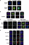
Shwachman-Diamond syndrome (SDS) is an autosomal recessively inherited disorder characterized by exocrine pancreatic insufficiency and bone marrow failure. The gene for this syndrome, SBDS, encodes a highly conserved novel protein. We characterized Shwachman-Bodian-Diamond syndrome (SBDS) protein expression and intracellular localization in 7 patients with SDS and healthy controls. As predicted by gene mutation, 4 patients with SDS exhibited no detectable full-length SBDS protein. Patient DF277, who was homozygous for the IVS2 + 2 T>C splice donor mutation, expressed scant levels of SBDS protein. Patient SD101 expressed low levels of SBDS protein harboring an R169C missense mutation. Patient DF269, who carried no detectable gene mutations, expressed wild-type levels of SBDS protein to add further support to the growing body of evidence for additional gene(s) that might contribute to the pathogenesis of the disease phenotype. The SBDS protein was detected in both the nucleus and the cytoplasm of normal control fibroblasts, but was particularly concentrated within the nucleolus. SBDS localization was cell-cycle dependent, with nucleolar localization during G1 and G2 and diffuse nuclear localization during S phase. SBDS nucleolar localization was intact in SD101 and DF269. The intranucleolar localization of SBDS provides further supportive evidence for its postulated role in rRNA processing.


Figures
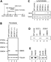
Figure 1.
Figure 1.
Patients with SDS express variable levels of SBDS protein. (A) Schematic diagram of SBDS mutations. The locations of the SBDS mutations identified in our patient series are diagramed (not drawn to scale). The SBDS gene is composed of 5 exons (depicted with boxes). The translated mRNA regions are shaded gray. (B) SBDS expression in fibroblasts. Bone marrow fibroblast protein lysates were analyzed by immunoblotting for SBDS protein (top). The SBDS genotypes of each cell line are indicated in parentheses. The blot was also probed for tubulin to ascertain equivalent protein loading (bottom). DF259 and DF307 harbored SBDS mutations predicted to lead to premature translation termination. The wild-type (WT) control (NMF-100) is a marrow fibroblast cell line derived from a healthy control patient. (C) SBDS expression in lymphoblasts. Lymphoblast protein lysates were analyzed by immunoblotting for SBDS protein (top) and tubulin (bottom). DF250, DF259, and DF260 carried SBDS mutations predicted to lead to premature translation termination. SD101 carried a missense (R169C) mutation on 1 SBDS allele. DF269 is derived from a patient with SDS who lacked any identified SBDS mutations. (D) Retroviral transduction of the SBDS cDNA restores SBDS protein expression. A lymphoblast (DF277.L) or fibroblast (DF259.F) SDS cell line was infected with a pBabe retrovirus containing a wild-type SBDS cDNA (lanes 3 and 5) or empty vector (lanes 2 and 4). Cell lysates were immunoblotted for SBDS protein. A normal lymphoblast control cell line (DF213) was run in lane 1 (WT control). (E) SBDS protein is expressed in a variety of tissues. Lysates from the indicated human cell lines were immunoblotted for SBDS protein expression. Caco2 indicates intestinal epithelial cell line; HepG2, hepatocellular carcinoma cell line; HL-60, myeloid leukemia cell line; U2OS, osteosarcoma cell line; CAPAN-1, pancreatic adenocarcinoma cell line.
Figure 2.
Figure 2.
SBDS localizes to the nucleolus. (A) Retroviral SBDS transduction restores SBDS expression by immunofluorescence. DF259.F fibroblasts infected with retrovirus containing empty vector (top) or SBDS cDNA (bottom) were fixed and probed with the anti-SBDS antibody followed by a FITC-labeled secondary antibody. The cells were counterstained with DAPI (4′, 6-diamino-2-phenylindole) to visualize the nuclei. SBDS was located throughout the cell but was particularly prominent in the region corresponding to the nucleolus. (B) Endogenous SBDS localizes to the nucleolus. HeLa cells were fixed and probed with both an anti-SBDS polyclonal antibody and an antinucleolin monoclonal antibody. SBDS was detected with an antirabbit FITC-labeled secondary antibody, and nucleolin was detected with an antimouse Texas-Red (TR) secondary antibody. Cells were visualized by standard fluorescence microscopy (top) or by confocal microscopy (bottom). (C) SBDS nucleolar localization is intact in SD101 fibroblasts. WT control fibroblasts (NMF-100) or SD101.F fibroblasts were analyzed by immunofluorescence for SBDS. Low power, × 63 magnification; high power, × 100 magnification. (D) SBDS localizes to the nucleolus in DF269 lymphoblasts and in myeloid cells. The indicated lymphoblast cell lines or the myeloid leukemia HL-60 cell lines were subjected to immunofluorescent staining for SBDS protein and nucleolin. Nuclei were counterstained with DAPI.
Figure 3.
Figure 3.
SBDS nucleolar localization is cell-cycle dependent. (A) HeLa cells were synchronized with a double thymidine block, then released for the times indicated. Cells were fixed and stained for SBDS. An aliquot of cells was also fixed for flow cytometry (right). Cells in late telophase are indicated with arrows. (B) SBDS protein levels remain constant throughout the cell cycle. HeLa cells synchronized with a double thymidine block were released for the indicated times. Cell lysates were immunoblotted for Fanconi anemia, complementation group D2 (FANCD2), SBDS, and tubulin.

References

    1. Shwachman H, Diamond LK, Oski FA, Khaw KT. The syndrome of pancreatic insufficiency and bone marrow dysfunction. J Pediatr. 1964;65: 645-663. - PubMed
    1. Bodian M, Sheldon W, Lightwood R. Congenital hypoplasia of the exocrine pancreas. Acta Paediatr. 1964;53: 282-293. - PubMed
    1. Ginzberg H, Shin J, Ellis L, et al. Segregation analysis in Shwachman-Diamond syndrome: evidence for recessive inheritance. Am J Hum Genet. 2000;66: 1413-1416. - PMC - PubMed
    1. Dror Y, Freedman MH. Shwachman-diamond syndrome. Br J Haematol. 2002;118: 701-713. - PubMed
    1. Smith OP. Shwachman-Diamond syndrome. Semin Hematol. 2002;39: 95-102. - PubMed

Publication types