. 2005 Oct 1;106(7):2366-74.
doi: 10.1182/blood-2004-10-4166. Epub 2005 Apr 28.

Chemokine up-regulation in SARS-coronavirus-infected, monocyte-derived human dendritic cells

Affiliations

Chemokine up-regulation in SARS-coronavirus-infected, monocyte-derived human dendritic cells

Helen K W Law et al. Blood. .

Abstract

Lymphopenia and increasing viral load in the first 10 days of severe acute respiratory syndrome (SARS) suggested immune evasion by SARS-coronavirus (CoV). In this study, we focused on dendritic cells (DCs) which play important roles in linking the innate and adaptive immunity. SARS-CoV was shown to infect both immature and mature human monocyte-derived DCs by electron microscopy and immunofluorescence. The detection of negative strands of SARS-CoV RNA in DCs suggested viral replication. However, no increase in viral RNA was observed. Using cytopathic assays, no increase in virus titer was detected in infected DCs and cell-culture supernatant, confirming that virus replication was incomplete. No induction of apoptosis or maturation was detected in SARS-CoV-infected DCs. The SARS-CoV-infected DCs showed low expression of antiviral cytokines (interferon alpha [IFN-alpha], IFN-beta, IFN-gamma, and interleukin 12p40 [IL-12p40]), moderate up-regulation of proinflammatory cytokines (tumor necrosis factor alpha [TNF-alpha] and IL-6) but significant up-regulation of inflammatory chemokines (macrophage inflammatory protein 1alpha [MIP-1alpha], regulated on activation normal T cell expressed and secreted [RANTES]), interferon-inducible protein of 10 kDa [IP-10], and monocyte chemoattractant protein 1 [MCP-1]). The lack of antiviral cytokine response against a background of intense chemokine up-regulation could represent a mechanism of immune evasion by SARS-CoV.


Figures
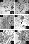
Figure 1.
Figure 1.
Electron microscopy of SARS-CoV–infected human DCs. Negative-contrast thin-section transmission electron micrograph of SARS-CoV–infected human DCs showed virus (black arrowheads) binding (A) and uptake (B) at 0 hours after infection. At 6 hours, 12 hours, and 24 hours after infection, virus particles were detected in endosomes (C) and cytoplasm (D) but not in the Golgi apparatus (E). No virus budding was observed (F) in all cells examined. Images are representative of SARS-CoV–infected human DCs from 3 independent adult or CB donors.
Figure 2.
Figure 2.
Immunofluorescence assay for SARS-CoV detection in human DCs. Mock-infected human DCs were included as a control (A). Positive immunofluorescence staining was detected in human immature and mature DCs at 12 hours (B) and 24 hours (C) after infection with SARS-CoV (MOI = 1). Confocal microscopy showed positive staining in the cytoplasm of DCs (D). Images are representative of immature and mature DCs from 11 independent adult or CB donors.
Figure 3.
Figure 3.
Viral gene expression in SARS-CoV–infected human adult immature DCs by quantitative RT-PCR. Both negative (A) and positive strands (B) of SARS-CoV Replicase 1b mRNA were detected in SASR-CoV–infected adult immature DCs at 3 hours, 9 hours, 24 hours, day 3, and day 6 after infection (MOI = 1). Similar pattern of decreased viral gene expression was detected for the negative and positive strands in all 3 cases.
Figure 4.
Figure 4.
Active caspase-3 assay for apoptosis in human adult immature DCs. Comparing with mock-infected adult immature DCs, no significant induction of active caspase-3–positive cells was observed at 6 hours, 12 hours, and 24 hours after infection (n = 4; P > .05). Data are shown as mean ± SEM of DCs from 4 independent donors. The percentage of active caspase-3–positive Jurkat cells in the positive control at 3 hours and 24 hours after addition of anti-Fas antibodies were 15% and 35%, respectively.
Figure 5.
Figure 5.
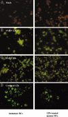
Flow cytometry analysis of cell-surface molecule expression on human adult DCs. Mock- (A) or SARS-CoV–infected (B) adult immature DCs (MOI = 1) were harvested at 48 hours after infection and stained for flow cytometry analysis. Surface staining is shown by filled histogram, and isotype control is marked by the dotted line. SARS-CoV alone did not up-regulate the expression of CD83, CD86, MHC class I, and MHC class II. However, SARS-CoV–infected cells can be stimulated by LPS (10 μg/mL; thick line) to up-regulate the expression of these molecules to similar levels as in the mock-infected controls. Data shown are representative of adult immature DCs from 5 independent donors. FSC indicates forward scatter; SSC, side scatter.
Figure 6.
Figure 6.
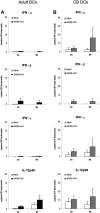
Antiviral cytokine gene expression profile of SARS-CoV–infected human immature DCs by quantitative RT-PCR. Antiviral cytokine mRNA concentrations in adult (A) and CB (B) immature cells were assayed at 3 hours and 9 hours after infection with SARS-CoV (MOI = 1). Mock-infected cells were included as negative control. The concentrations were normalized to those of β-actin mRNA in the corresponding sample. There were low expressions of IFN-α, IFN-β, IFN-γ, and IL-12p40 genes in SARS-CoV–infected DCs. Data are shown as mean ± SEM (adult n = 7; CB n = 5).
Figure 7.
Figure 7.
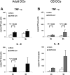
Proinflammatory cytokine gene expression profile of SARS-CoV–infected human immature DCs by quantitative RT-PCR. Proinflammatory cytokine mRNA concentrations in adult (A) and CB (B) immature cells were assayed at 3 hours and 9 hours after infection with SARS-CoV (MOI = 1). Mock-infected cells were included as negative control. The concentrations were normalized to those of β-actin mRNA in the corresponding sample. There were moderate up-regulation of TNF-α and IL-6 expression in SARS-CoV–infected DCs. Data are shown as mean ± SEM (adult n = 7; CB n = 5; *P < .05).
Figure 8.
Figure 8.
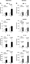
Chemokine gene expression profile of SARS-CoV–infected human immature DCs by quantitative RT-PCR. Chemokine mRNA concentrations in adult (A) and CB (B) immature cells were assayed at 3 hours and 9 hours after infection with SARS-CoV (MOI = 1). Mock-infected cells were included as negative control. The concentrations were normalized to those of β-actin mRNA in the corresponding sample. There was significant up-regulation of MIP-1α, RANTES, IP-10, and MCP-1 in SARS-CoV–infected DCs. Data are shown as mean ± SEM (adult n = 7; CB n = 5; *P < .05).

Similar articles

Cited by

References

    1. Lai, MMC, Holmes KV. Coronaviridae: the viruses and their replication. In: Knipe DM, Howley PM, eds. Fields' virology. Philadelphia, PA: Lippincott Williams & Wilkins; 2001: 1163-1185.
    1. Choi KW, Chau TN, Tsang O, et al. Outcomes and prognostic factors in 267 patients with severe acute respiratory syndrome in Hong Kong. Ann Intern Med. 2003;139: 715-723. - PubMed
    1. Peiris JS, Lai ST, Poon LL, et al. Coronavirus as a possible cause of severe acute respiratory syndrome. Lancet. 2003;361: 1319-1325. - PMC - PubMed
    1. Peiris JS, Chu CM, Cheng VC, et al. Clinical progression and viral load in a community outbreak of coronavirus-associated SARS pneumonia: a prospective study. Lancet. 2003;361: 1767-1772. - PMC - PubMed
    1. Drosten C, Gunther S, Preiser W, et al. Identification of a novel coronavirus in patients with severe acute respiratory syndrome. N Engl J Med. 2003; 348: 1967-1976. - PubMed

Publication types

MeSH terms