. 2005 May 4;24(9):1686-95.
doi: 10.1038/sj.emboj.7600647. Epub 2005 Apr 7.

Association of Csk to VE-cadherin and inhibition of cell proliferation

Affiliations

Association of Csk to VE-cadherin and inhibition of cell proliferation

Ulf Baumeister et al. EMBO J. .

Abstract

Vascular endothelial cadherin (VE-cadherin) mediates contact inhibition of cell growth in quiescent endothelial cell layers. Searching for proteins that could be involved in VE-cadherin signaling, we found the cytosolic C-terminal Src kinase (Csk), a negative regulator of Src family kinases. We show that Csk binds via its SH2 domain to the phosphorylated tyrosine 685 of VE-cadherin. VE-cadherin recruits Csk to cell contacts and both proteins can be co-precipitated from cell lysates of transfected cells and endothelial cells. Association of VE-cadherin and Csk in endothelial cells increased with increasing cell density. CHO cells expressing the tyrosine replacement mutant VE-cadherin-Y685F grow to higher cell densities than cells expressing wild-type VE-cadherin. Overexpression of Csk in these cells under an inducible promoter inhibits cell proliferation in the presence and absence of VE-cadherin, but not in the presence of VE-cadherin-Y685F. Reduction of Csk expression by RNA interference enhances endothelial cell proliferation. Our results suggest that the phosphorylated tyrosine residue 685 of VE-cadherin and probably the binding of Csk to this site are involved in inhibition of cell growth triggered by cell density.


Figures

Figure 1
Figure 1
Designation of recombinant forms of Csk and VE-cadherin. (A) All forms of Csk carried a Myc tag at the N-terminus. Besides the wt form (Csk-wt), two mutants were generated, one with arginine 107 in the SH2 domain replaced by lysine (Csk-R107K) and the other with lysine 222 in the kinase domain replaced by an arginine (Csk-K222R). The truncated form Csk57–204 was coded by the cDNA fragment picked in the yeast two-hybrid screen with a VE-cadherin bait construct. (B) The following forms of VE-cadherin are depicted: wt form of VE-cadherin (VE-cad-wt), a tyrosine replacement mutant of full-length VE-cadherin (VE-cad-Y685F), a fragment of the cytoplasmic tail of VE-cadherin (VE-cad621–764), which was inserted in the yeast two-hybrid bait construct (see Materials and methods for details) and two GST fusion proteins containing the same part of the cytoplasmic tail of VE-cadherin as the bait construct. One of the two GST fusion proteins contained the Y685F point mutation. The binding sites for p120 and β-catenin are demarcated in analogy to the binding sites for E-cadherin.
Figure 2
Figure 2
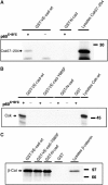
In vitro-translated Csk can be precipitated with a GST-VE-cadherin fusion protein. Either the Csk57–204 fragment identified in the yeast two-hybrid screen (A) or full-length Csk (Csk-wt) (B) was synthesized in a coupled in vitro transcription/translation reaction and precipitation was tested with the indicated GST-VE-cadherin (see Figure 1) and the GST-N-cadherin (GST-N-cad) fusion proteins. An aliquot of the translation reaction is shown in the right lane. The intactness of the GST-cadherin fusion proteins was tested in pull-down experiments with in vitro-translated β-catenin (C). Molecular mass markers are indicated on the right.
Figure 3
Figure 3
Csk can be co-immunoprecipitated together with VE-cadherin from CHO cell lysates. (A) CHO cells stably cotransfected with inducible Csk and either wt-VE-cadherin or the tyrosine replacement mutant (as indicated above) were subjected to immunoprecipitations with polyclonal preimmune antibodies (preimmune) or affinity-purified anti-VE-cadherin antibodies, and precipitates were blotted with antibodies against Csk (upper panel) or VE-cadherin (middle panel). Aliquots of cell lysates were directly analyzed in immunoblots for expression of Csk (bottom panel). Induction of exogenous Csk expression (Induction) is indicated above. Note that transfected, Myc-tagged Csk has a slightly higher apparent molecular weight than endogenous Csk. The asterisk marks weakly co-precipitated endogenous Csk. (B) CHO cells stably cotransfected with inducible Csk and constitutively expressed wt-VE-cadherin were subjected to immunoprecipitations with anti-Myc-tag antibodies (α-Myc) and precipitates were blotted with antibodies against VE-cadherin (upper panel) or Csk (middle panel). Aliquots of cell lysates were directly analyzed in immunoblots for expression of VE-cadherin (bottom panel). Molecular mass markers are indicated on the right.
Figure 4
Figure 4
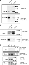
Endogenously expressed Csk can be co-precipitated with VE-cadherin from lysates of endothelial cells and association increases with cell density. (A, C) Mouse bEnd.3 endothelioma cells and (B) HUVECs were subjected to immunoprecipitations with (A, C) affinity-purified polyclonal anti-mouse VE-cadherin antibodies (αVE-cad) and preimmune antibodies (ctr). (B) mAb against human VE-cadherin (αVE-cad) and an irrelevant class-matched mAb (ctr). Precipitates were analyzed in immunoblots with anti-Csk antibodies and the same filters were re-analyzed with anti-VE-cadherin antibodies (as indicated). Expression levels of Csk and VE-cadherin were analyzed by immunoblotting cell lysates (lysates). (C) Two different cell densities were plated by splitting confluent monolayers 1:2 or 1:5. Cell lysates containing identical protein amounts were analyzed. Molecular mass markers are indicated on the right.
Figure 5
Figure 5
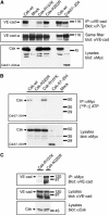
Coexpression of Csk with VE-cadherin in COS-7: upregulation of tyrosine phosphorylation requires SH2 domain, but not kinase domain. (A) COS-7 cells were transiently cotransfected with VE-cadherin and various recombinant forms of Csk as indicated above. Cell lysates were immunoprecipitated with anti-VE-cadherin antibodies and precipitates were analyzed in immunoblots first with anti-phospho-tyrosine antibodies (αP-Tyr) and then (on the same filter) with anti-VE-cadherin antibodies (αVE-cad). Expression of the various forms of Csk was controlled in immunoblots of cell lysates (bottom panel). Note that the Csk-SH2 domain mutant (Csk-R107K) caused strongly reduced tyrosine phosphorylation of VE-cadherin, whereas the Csk kinase mutant (Csk-K222R) was not impaired in its ability to stimulate tyrosine phosphorylation of VE-cadherin. (B) Analysis of the kinase activity of Csk mutants. Various recombinant forms of Csk were expressed in COS-7 cells, immunoprecipitated with anti-Myc-tag antibodies and the kinase activity of the precipitated proteins was analyzed by incubation with [32P-γ]ATP, followed by gel electrophoresis and autoradiography (upper panel). Efficiency of immunoprecipitations was controlled by analyzing immunoblots of equivalent aliquots of the precipitates with anti-Myc-tag antibodies (lower panel). Note that no kinase activity was detectable for the kinase point mutation Csk-K222R. (C) Analysis of the association of the two Csk point mutants with VE-cadherin: COS-7 cells were transfected with VE-cadherin and in addition either with the Csk-R107K SH2 domain mutant or with the Csk-K222R kinase mutant and cell lysates were immunoprecipitated with anti-Myc-tag antibodies. Precipitates were analyzed in immunoblots with anti-VE-cadherin antibodies (upper panel). Expression levels of VE-cadherin and Csk were tested in immunoblots of cell lysates (middle and bottom panels). Note that binding of the SH2 domain mutant to VE-cadherin was strongly but not completely inhibited. Molecular mass markers are indicated on the right.
Figure 6
Figure 6
VE-cadherin recruits Csk to cell contacts. CHO cells stably transfected with Csk under an inducible promoter and either coexpressing wt-VE-cadherin (upper row) or the tyrosine mutant VE-cad-Y685F (bottom row) were analyzed by indirect immunofluorescence staining for the localization of Csk (red, left column) and VE-cadherin (green, middle column). The merge is shown in the right column. In all cases, Csk expression was induced. Bar=16 μm.
Figure 7
Figure 7
Induction of Csk in transfected CHO cells slows down cell proliferation only if wt-VE-cadherin is coexpressed. CHO cells expressing inducible wt form of Csk (Csk-wt) or the kinase mutant of Csk (Csk-mt) were cotransfected with wt form of VE-cadherin (VE-cad-wt), or VE-cadherin-Y685F (VE-cad-Y685F) or without VE-cadherin (without VE-cad), and were analyzed for [3H]thymidine incorporation. In each case, two different transfected clones were analyzed. For each clone, [3H]thymidine incorporation of cells not induced for Csk expression was set as 100% (black bar). Measurements were carried out with cells at early confluence, resembling the stage between 72 and 96 h growth in Figure 8. At this stage, [3H]thymidine incorporation of wt-VE-cadherin and mt-VE-cadherin cells in the absence of Csk induction was similar (within a range of 15%, not shown), in agreement with the results in Figure 8.
Figure 8
Figure 8
CHO cells expressing mutant VE-cadherin-Y685-F grow to higher cell densities than CHO cells expressing wt-VE-cadherin. Identical cell numbers of the two CHO clones 20.9 and 29.5 each transfected with wt-VE-cadherin and inducible Csk and the two CHO clones 3.30 and 2.24 each transfected with mutant VE-cadherin-Y685-F and inducible Csk were plated and cells were harvested and counted at the indicated time points (in hours). Cell densities are given on the left as cells per cm2. Note that these cells were not induced for exogenous Csk expression and the effects shown are based on endogenous Csk expression levels.
Figure 9
Figure 9
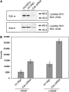
Downregulation of Csk by siRNA increases cell proliferation of HUVECs. (A) Immunoblot analysis for Csk expression of lysates of HUVECs that had been transfected with siRNA against an irrelevant mouse antigen (ctr. oligo) or with siRNA targeting Csk (Csk siRNA) or not transfected (HUVECs). Results are shown at 24 and 48 h after transfection (as indicated). (B) HUVECs were either transfected with siRNA against an irrelevant mouse antigen (ctr. oligo) or with a Csk-specific siRNA (Csk siRNA) and incorporation of [3H]thymidine into cells was measured either in the absence (−VEGF) or presence (+VEGF) of added VEGF.

References

    1. Abu-Ghazaleh R, Kabir J, Jia H, Lobo M, Zachary I (2001) Src mediates stimulation by vascular endothelial growth factor of the phosphorylation of focal adhesion kinase at tyrosine 861, and migration and anti-apoptosis in endothelial cells. Biochem J 360: 255–264 - PMC - PubMed
    1. Borges E, Tietz W, Steegmaier M, Moll T, Hallmann R, Hamann A, Vestweber D (1997) P-selectin glycoprotein ligand-1 (PSGL-1) on T helper 1 but not on T helper 2 cells binds to P-selectin and supports migration into inflamed skin. J Exp Med 185: 573–578 - PMC - PubMed
    1. Breier G, Breviario F, Caveda L, Berthier R, Schnurch H, Gotsch U, Vestweber D, Risau W, Dejana E (1996) Molecular cloning and expression of murine vascular endothelial-cadherin in early stage development of cardiovascular system. Blood 87: 630–641 - PubMed
    1. Cao MY, Huber M, Beauchemin N, Famiglietti J, Albelda SM, Veillette A (1998) Regulation of mouse PECAM-1 tyrosine phosphorylation by the Src and Csk families of protein-tyrosine kinases. J Biol Chem 273: 15765–15772 - PubMed
    1. Caveda L, Martin-Padura I, Navarro P, Breviario F, Corada M, Gulino D, Lampugnani MG, Dejana E (1996) Inhibition of cultured cell growth by vascular endothelial cadherin (cadherin-5/VE-cadherin). J Clin Invest 98: 886–893 - PMC - PubMed

MeSH terms