. 2005 Aug 15;390(Pt 1):67-75.
doi: 10.1042/BJ20050414.

Roles of 14-3-3 and calmodulin binding in subcellular localization and function of the small G-protein Rem2

Affiliations

Roles of 14-3-3 and calmodulin binding in subcellular localization and function of the small G-protein Rem2

Pascal Béguin et al. Biochem J. .

Erratum in

  • Biochem J. 2005 Nov 1;391(Pt 3):712

Abstract

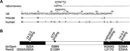
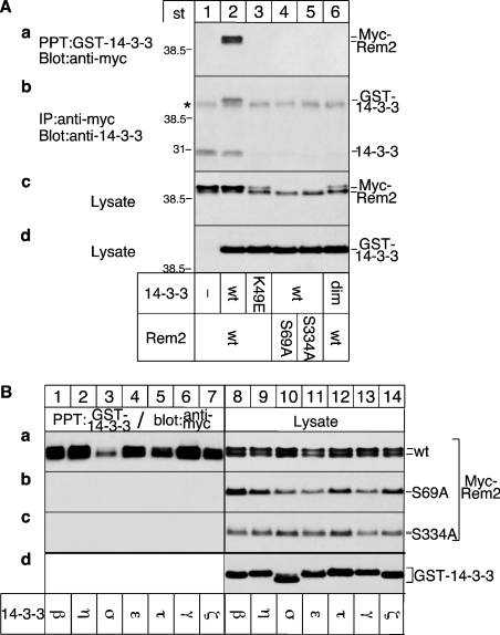
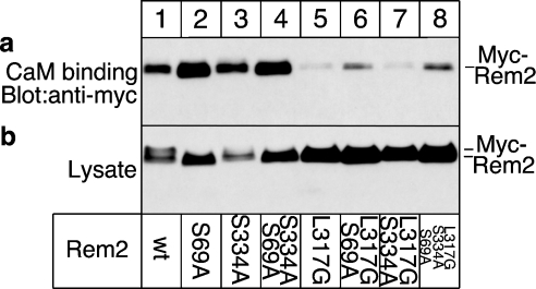
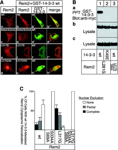
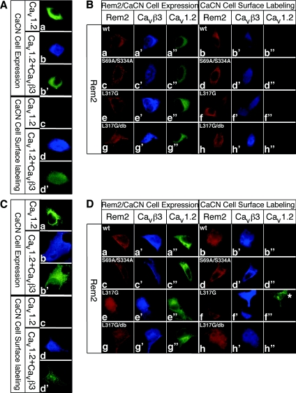
kir/Gem, Rad, Rem and Rem2 comprise the RGK (Rad/Gem/kir) family of Ras-related small G-proteins. Two important functions of RGK proteins are the regulation of the VDCC (voltage-dependent Ca2+ channel) activity and cell-shape remodelling. RGK proteins interact with 14-3-3 and CaM (calmodulin), but their role on RGK protein function is poorly understood. In contrast with the other RGK family members, Rem2 has been reported to bind neither 14-3-3 nor induce membrane extensions. Furthermore, although Rem2 inhibits VDCC activity, it does not prevent cell-surface transport of Ca2+ channels as has been shown for kir/Gem. In the present study, we re-examined the functions of Rem2 and its interaction with 14-3-3 and CaM. We show that Rem2 in fact does interact with 14-3-3 and CaM and induces dendrite-like extensions in COS cells. 14-3-3, together with CaM, regulates the subcellular distribution of Rem2 between the cytoplasm and the nucleus. Rem2 also interacts with the beta-subunits of VDCCs in a GTP-dependent fashion and inhibits Ca2+ channel activity by blocking the alpha-subunit expression at the cell surface. Thus Rem2 shares many previously unrecognized features with the other RGK family members.


Figures

Figure 1
Figure 1. Identification of 14-3-3-binding sites in Rem2
(A) N-terminal amino acid sequence of full-length rat Rem2 and a truncated form (AF084464) [21] lacking the N-terminal 69 amino acids. The nucleotide sequence of the cDNA encoding the Rem2 and the truncated form indicate that the omission of a C in the truncated form resulted in an in-frame stop codon upstream of Met70, erroneously assigned as the initiation methionine. The N-terminal amino acid sequence of Rem2 for rat, mouse and human is aligned, showing that Met1 is conserved. An in-frame stop codon is located upstream of Met1 (results not shown). Amino acids are shown in one-letter code and residues conserved between species are indicated with a dashed line. (B) Domain structure of Rem2. The Ras-like core domain (white bar), N- and C-terminal extensions (black bars) and the location of the 14-3-3, CaM (white circles) and GTP (black circle)-binding sites are shown. Mutations that affect the different binding sites are indicated, with non-functional putative 14-3-3-binding sites in brackets.
Figure 2
Figure 2. Identification and characterization of 14-3-3 binding sites in Rem2
(A) Identification of 14-3-3-binding sites. (a) Cells were co-transfected with cDNAs for wt or mutated Myc-Rem2 and GST–14-3-3, GST–14-3-3 K49E ζ isoforms or a dimerization defective mutant (dim). GST–14-3-3 proteins were precipitated and associated Rem2 detected by Western blotting using Myc antibodies. (b) Cells were co-transfected with cDNAs for wt or mutated Myc-Rem2 and GST–14-3-3, GST–14-3-3 K49E or a dimerization defective mutant (dim). Rem2 was immunoprecipitated and associated GST–14-3-3 and endogenous 14-3-3 were detected by Western blotting using 14-3-3 antibodies. The IgG heavy chain, migrating just below the GST–14-3-3 in (b), is marked by an *. (c, d) Expression levels. Cell lysates were blotted with Myc (c) or GST (d) antibodies to monitor the expression level of Myc–Rem or GST–14-3-3 respectively; st, protein markers of known molecular mass. (B) Association of Rem2 with 14-3-3 isoforms. (a–c, lanes 1–7) Cells were co-transfected with cDNAs for the different GST–14-3-3 isoforms and wt or mutated Myc–Rem proteins. GST–14-3-3 proteins were precipitated and associated RGK proteins were detected by Western blotting using a Myc antibody. (a–c, lanes 8–14) Cell lysates were blotted with Myc antibodies to monitor Rem2-expression levels. (d) One representative example of cell lysates blotted with GST antibody to monitor expression levels of the GST–14-3-3 isoforms.
Figure 3
Figure 3. Binding of CaM to Rem2
(a) Cells were transfected with cDNA for wt or mutated Myc–Rem2. Cell homogenates were incubated with CaM beads and bound Myc–Rem2 detected by Western blotting using Myc antibody. (b) Cell lysates were blotted with Myc antibodies to monitor Rem2 expression levels.
Figure 4
Figure 4. 14-3-3 and CaM regulate the subcellular distribution of Rem2, and Rem2 mediates changes to cell morphology
(A) COS-1 cells were transfected with cDNAs for wt or mutated Rem2, either alone or together with GST–14-3-3. Cells were processed for immunofluorescence microscopy using Myc and GST antibodies to label Rem2 (a–h; red) and GST–14-3-3 (b′, d′ f′ and h′; green) respectively. Areas of co-localization are in yellow in the merged images (b″, d″, f″ and h″); db, double mutant. (B) Rem2 mutants defective in CaM binding associate with 14-3-3. Cells were co-transfected with cDNAs for mutated Myc–Rem2 and GST–14-3-3 or GST–14-3-3 K49E. (a) GST–14-3-3 proteins were precipitated and the associated Myc–Rem2 detected by Western blotting using Myc antibody. Cell lysates were blotted with Myc (b) or GST (c) antibodies to monitor Rem2 or GST–14-3-3 expression levels respectively. (C) Quantification of the 14-3-3 mediated cytosolic relocalization of Rem2. Transfected cells (150–200) were randomly selected and analysed in 3–5 independent experiments. The fraction of cells showing efficient (black bars), partial (grey bar) or no (white bars) nuclear clearance is plotted.
Figure 5
Figure 5. Nucleotide-dependent binding of Rem2 to the β-subunit of VDCCs and down-regulation of Ca2+ channel activity
(A) Pull down. Cells were transfected with cDNAs Myc–Rem2, Rem2 S129N or Rem2 L317G. Cell homogenates were incubated with immobilized recombinant GST–Cavβ3 and associated Rem2 was detected by Western blotting using Myc antibody. Recombinant GST served as a control (lane 1). Cell lysates were blotted with Myc antibodies to monitor Rem2 expression levels (lanes 5–7). (B) Electrophysiology. PC-12 cells were co-transfected with a GFP (green fluorescent protein) plasmid and cDNAs for wt or mutated Rem2 either with or without 14-3-3. GFP-positive cells were selected for electrophysiology and the average of the maximal current detected at +20 mV for endogenous Ca2+ channels was measured. For each condition, 9–16 independent experiments were carried out. An example of the IV relationship of Ca2+ channels in PC-12 cells is shown below. A, Ampere; F, Farad. Cells transfected only with the GFP cDNA served as a control.
Figure 6
Figure 6. Rem2 blocks cell-surface expression of VDCC α-subunits in PC-12 and HEK-293T cells
(A, C) The β-subunit facilitates surface expression of the α-subunit. PC-12 (A) or HEK-293T (C) cells were transfected with a cDNA for Cav1.2 carrying an extracellular HA tag or with an IRES-based vector carrying the cDNAs for HA–Cav1.2 and FLAG–Cavβ3 subunits. Cells were fixed, permeabililzed and processed for immunofluorescence microscopy using HA and FLAG antibodies to detect Cav1.2 (green) and Cavβ3 (blue) respectively (CaCN cell expression). Alternatively, live cells were first incubated with HA antibodies to selectively label surface exposed Cav1.2 before the fixation, permeabilization and labelling with FLAG antibodies (CaCN cell-surface labelling). (B, D) Effect of Rem2 on cell-surface expression of α-subunits. PC-12 (B) or HEK-293T (D) cells were transfected with cDNAs for wt or mutated Myc–Rem2 together with an IRES-based vector carrying the cDNAs for HA–Cav1.2 and FLAG–Cavβ3. Cells were fixed, permeabililzed and processed for immunofluorescence microscopy using Myc, HA and FLAG antibodies to detect Rem2 (red), Cav1.2 (green) and Cavβ3 (blue) respectively (Rem2/CaCN cell expression). Alternatively, live cells were first incubated with HA antibodies to selectively label surface exposed Cav1.2 before the fixation, permeabiliziation and labelling with Myc and FLAG antibodies (CaCN cell-surface labelling). db: double mutant with both 14-3-3-binding sites mutated; * in (D f″) indicates a cell that does not express Rem2 and consequently Cav1.2 is expressed at the cell surface. Independent experiments (4 or 5) were performed and 20–30 cells expressing the α- and β-CaCN subunits and the Rem2 proteins were analysed.

References

    1. Maguire J., Santoro T., Jensen P., Siebenlist U., Yewdell J., Kelly K. Gem: an induced, immediate early protein belonging to the Ras family. Science. 1994;265:241–244. - PubMed
    1. Cohen L., Mohr R., Chen Y. Y., Huang M., Kato R., Dorin D., Tamanoi F., Goga A., Afar D., Rosenberg N., et al. Transcriptional activation of a ras-like gene (kir) by oncogenic tyrosine kinases. Proc. Natl. Acad. Sci. U.S.A. 1994;91:12448–12452. - PMC - PubMed
    1. Reynet C., Kahn C. R. Rad: a member of the Ras family overexpressed in muscle of type II diabetic humans. Science. 1993;262:1441–1444. - PubMed
    1. Finlin B. S., Andres D. A. Rem is a new member of the Rad- and Gem/Kir Ras-related GTP-binding protein family repressed by lipopolysaccharide stimulation. J. Biol. Chem. 1997;272:21982–21988. - PubMed
    1. Finlin B. S., Shao H., Kadono-Okuda K., Guo N., Andres D. A. Rem2, a new member of the Rem/Rad/Gem/Kir family of Ras-related GTPases. Biochem. J. 2000;347:223–231. - PMC - PubMed

Publication types

MeSH terms