. 2005 Jun;17(6):1801-14.
doi: 10.1105/tpc.105.031419. Epub 2005 Apr 29.

Destination-selective long-distance movement of phloem proteins

Affiliations

Destination-selective long-distance movement of phloem proteins

Koh Aoki et al. Plant Cell. 2005 Jun.

Abstract

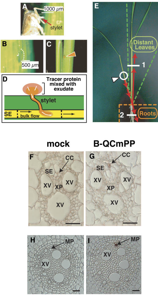
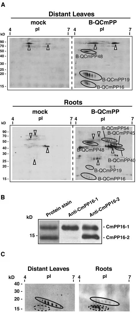
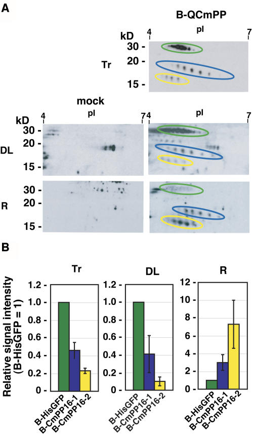
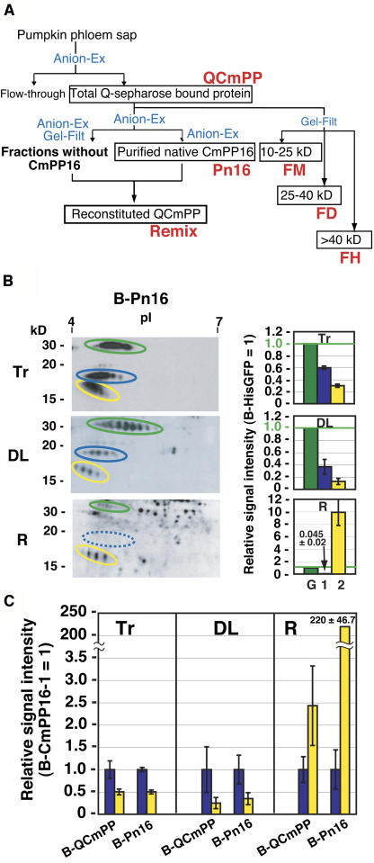
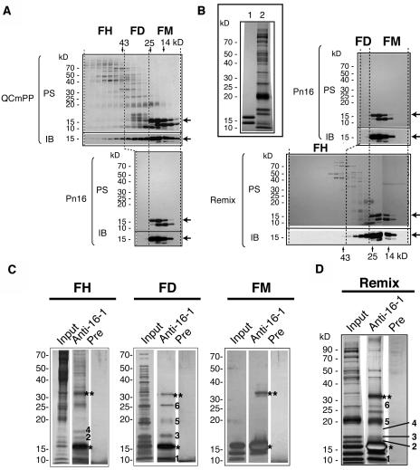
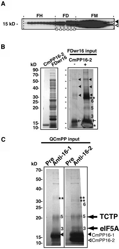
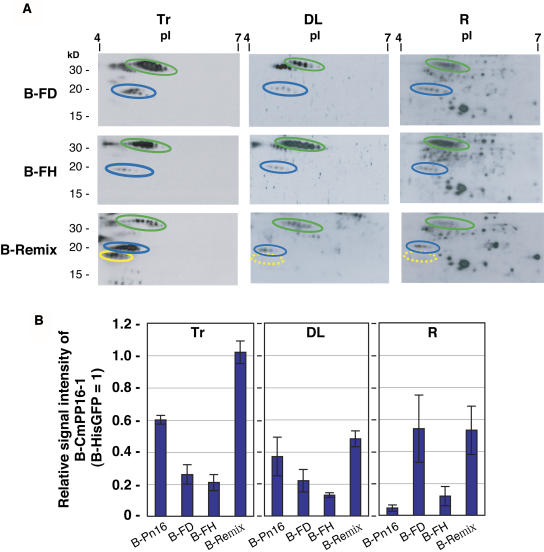
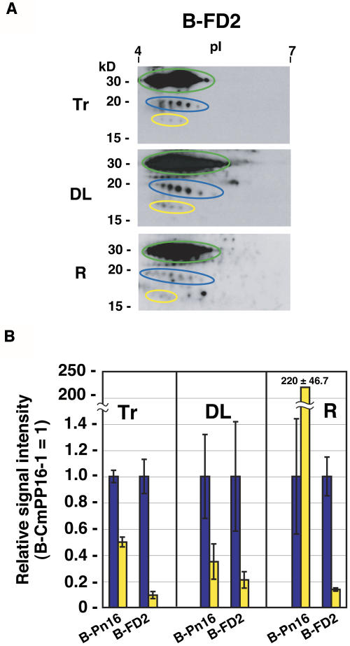
The phloem macromolecular transport system plays a pivotal role in plant growth and development. However, little information is available regarding whether the long-distance trafficking of macromolecules is a controlled process or passive movement. Here, we demonstrate the destination-selective long-distance trafficking of phloem proteins. Direct introduction, into rice (Oryza sativa), of phloem proteins from pumpkin (Cucurbita maxima) was used to screen for the capacity of specific proteins to move long distance in rice sieve tubes. In our system, shoot-ward translocation appeared to be passively carried by bulk flow. By contrast, root-ward movement of the phloem RNA binding proteins 16-kD C. maxima phloem protein 1 (CmPP16-1) and CmPP16-2 was selectively controlled. When CmPP16 proteins were purified, the root-ward movement of CmPP16-1 became inefficient, suggesting the presence of pumpkin phloem factors that are responsible for determining protein destination. Gel-filtration chromatography and immunoprecipitation showed that CmPP16-1 formed a complex with other phloem sap proteins. These interacting proteins positively regulated the root-ward movement of CmPP16-1. The same proteins interacted with CmPP16-2 as well and did not positively regulate its root-ward movement. Our data demonstrate that, in addition to passive bulk flow transport, a destination-selective process is involved in long-distance movement control, and the selective movement is regulated by protein-protein interaction in the phloem sap.


Figures

Figure 1.
Figure 1.
Strategy to Introduce Tracer Proteins into a Single Sieve Tube. (A) to (E) Outline of our experimental approach. (A) Brown leafhopper feeding on the phloem sap of rice. Cut the stylet of the insect by laser. (B) Phloem sap exudation through the cut stylet. (C) Tracer proteins were mixed with phloem exudates. (D) Tracer proteins entered the sieve element (SE) by diffusion. We frequently observed that the rate of exudation changed with time and could even go in the reverse (inward) direction. (E) Translocation pathway of tracer proteins. Incorporated tracer moved toward the basal end of the leaf and then moved to another leaf or to roots. The white arrowhead indicates a tracer application point. (F) to (I) Histochemical detection of tracer proteins in the distant organs. Leaf and root sections were obtained from positions 1 and 2, respectively, shown by the white lines in (E). No signal was detected in vascular tissues of leaf (H) or root (J) of plants treated with introduction buffer alone (mock). Tracer signal was detected in phloem tissues of distant leaves (I) and roots (K) of tracer-applied plants (B-QCmPP). The result was reproducible by sectioning three plants. CC, companion cell; MP, metaphloem tissue; SE, sieve element; XP, xylem parenchyma cell; XV, xylem vessel. Bars = 25 μm.
Figure 2.
Figure 2.
Profile of Tracer Proteins Detected in Rice Distant Organs. (A) Tracer proteins were detected by the combination of two-dimensional electrophoresis, on a 12% acrylamide gel, followed by ABC detection (2DE-ABC). Signals in the mock plant represent background signals from rice endogenous proteins (arrowheads). Several spot series were detected specifically in B-QCmPP-applied plants (B-QCmPP). Indicated spot series were subjected to internal peptide sequencing. BQCmPP48 was identified as SLW1, BQCmPP19 was identified as CmPP16-1, and BQCmPP16 was identified as CmPP16-2. BQCmPP54, BQCmPP45, and BQCmPP40 were left unidentified. Reproducible results were obtained from analysis of five plants. Forty micrograms of soluble protein was loaded per gel. (B) To confirm the presence of CmPP16 proteins in rice distant organs, anti-CmPP16-1 antibody and anti-CmPP16-2 antibody were prepared. The specificity of these antibodies was examined against phloem-purified CmPP16-1 and CmPP16-2. Anti-CmPP16-1 antibody reacted with CmPP16-1 50-fold more specifically than with CmPP16-2. Anti-CmPP16-2 antibody reacted with both CmPP16-1 and CmPP16-2 to the same extent. (C) Immunoblotting using anti-CmPP16-2 antibody revealed that B-CmPP19 (oval) and B-CmPP16 (broken oval) cross-reacted with the antibody, indicating that they were CmPP16-1 and CmPP16-2, respectively. The signal intensity ratio of the immunoreaction was similar to that of ABC detection. Proteins were run on 12% acrylamide gels. Note that immunodetection of tracer CmPP16 proteins was possible only when the biotin-derived signal in distant organs was very strong.
Figure 3.
Figure 3.
Semiquantitative Estimation of Long-Distance Movement of CmPP16 Proteins. (A) B-QCmPP tracer (Tr) was introduced, and then CmPP16 proteins were detected in rice distant leaves (DL) and roots (R) by 2DE-ABC detection. 2DE-ABC detection of mock-treated plants (mock) showed the background signals from rice endogenous proteins. B-HisGFP, B-CmPP16-1, and B-CmPP16-2 are indicated by green, blue, and yellow ovals, respectively. (B) Comparison of B-HisGFP (green bars), B-CmPP16-1 (blue bars), and B-CmPP16-2 (yellow bars) signals in tracer, distant leaves, and roots. The graph shows the signal intensity ratio of B-CmPP16-1 or B-CmPP16-2 to B-HisGFP. Values represent means ± se of three independent plants.
Figure 4.
Figure 4.
Long-Distance Movement of Purified CmPP16 Proteins. (A) Fractionation of pumpkin phloem sap protein by anion-exchange chromatography (Anion-Ex) and gel-filtration chromatography (Gel-Filt). Crude phloem sap was first loaded onto an anion-exchange column, and then total bound proteins were designated QCmPP. QCmPP was further fractionated by either Anion-Ex or Gel-Filt. Anion-Ex–purified CmPP16 was designated purified native CmPP16 (Pn16). All other Anion-Ex–eluted fractions free from CmPP16 protein were pooled and designated fraction without CmPP16. In reconstitution (shown in Figure 5B), Pn16 and fraction without CmPP16 were mixed to give reconstituted phloem proteins (Remix). Gel-Filt elution was subdivided into FM, FD, and FH according to molecular mass. (B) Biotinylated Pn16 (B-Pn16; Tr) was introduced into a rice sieve tube, and then the movement of B-HisGFP (green ovals), B-CmPP16-1 (blue ovals), and B-CmPP16-2 (yellow ovals) was detected in distant leaves (DL) and roots (R) by 2DE-ABC. In the root, B-CmPP16-1 signal was under the detection limit (blue broken oval). At right, relative signal intensities of B-CmPP16-1 (bar 1) and B-CmPP16-2 (bar 2) to B-HisGFP (bar G) are shown. (C) Comparison of relative signal intensities of B-CmPP16-2 (yellow bars) to B-CmPP16-1 (blue bars) in B-QCmPP and B-Pn16 introductions. Values represent means ± se of the signal intensity ratios from six independent plants. In the root, the [B-CmPP16-2/B-CmPP16-1] ratio increased significantly when B-Pn16 tracer was introduced. Values for B-QCmPP introduction were calculated from the relative signal intensity shown in Figure 3B.
Figure 5.
Figure 5.
CmPP16-1 Interacts with Specific Pumpkin Phloem Sap Proteins. (A) and (B) Comparison of gel-filtration profiles of QCmPP (top gel) and Pn16 (bottom gel) (A) and Pn16 (top gel) and Remix (bottom gel) (B). The inset in (B) shows protein profiles of Pn16 (lane 1) and a fraction without CmPP16 proteins (lane 2). Protein samples were run on a 12.5% acrylamide gel. In each gel, the top images show protein stain by Coomassie Brilliant Blue (PS) and the bottom images show immunoblotting using anti-CmPP16-1 antibody (IB). Arrows indicate CmPP16-1. Gel-filtration elution fractions were grouped into FM (10 to 25 kD), FD (25 to 40 kD), and FH (>40 kD). In QCmPP, CmPP16-1 was eluted in FM, FD, and FH. In Pn16, most of the CmPP16-1 was eluted in FM. (C) and (D) Immunoprecipitation assay using anti-CmPP16-1 antibody against QCmPP-derived FH, FD, and FM (C) and Remix fraction (D). Input proteins were immunoprecipitated using either anti-CmPP16-1 IgG (Anti-16-1) or preimmune IgG (Pre). Protein samples were run on a 12.5% acrylamide gel and silver-stained. The numbered bands represent coimmunoprecipitated proteins. Asterisks and double asterisks indicate CmPP16-1 monomer and CmPP16-1 dimer, respectively.
Figure 6.
Figure 6.
CmPP16-2 Interacts with the Same Pumpkin Phloem Sap Proteins as CmPP16-1. (A) Gel-filtration fractions of QCmPP were immunoblotted using anti-CmPP16-2 antibody (which cross-reacts with CmPP16-1 and CmPP16-2 equally well). Black and white arrowheads indicate CmPP16-1 and CmPP16-2, respectively. Small white arrowheads in FD indicate CmPP16-2 present in FD. CmPP16-2 was present in FM (monomer) and FD but not in FH. (B) Phloem-purified CmPP16-2 and FD with reduced amount of CmPP16 (FDwr16) were prepared separately (left). Coimmunoprecipitation was performed to isolate CmPP16-2–interacting protein from FD using anti-CmPP16-2 antibody (right). The same amount of FDwr16 (300 μg) was applied to the anti-CmPP16-2 antibody column with (lane +, right) or without (lane −, right) the addition of CmPP16-2 (30 μg). Samples were run on a 2.5% acrylamide gel and silver-stained. Asterisks and double asterisks indicate CmPP16-2 monomer and CmPP16-2 dimer, respectively. Arrowheads indicate the nonspecifically precipitated FDwr16 proteins. Arrows indicate residual CmPP16-1 in FDwr16 captured by anti-CmPP16-2 antibody. Bands 3, 5, and 6 were identical to those in Figure 5C. (C) Immunoprecipitation of QCmPP input using anti-CmPP16-1 IgG (Anti-16-1), anti-CmPP16-2 IgG (Anti-16-2), and preimmune IgG (Pre). Bands 3 and 5 were coimmunoprecipitated from unfractionated QCmPP. A faint signal of band 6 was detected in the Anti-16-2 lane but not in the Anti-16-1 lane. Double asterisks indicate dimers of CmPP16-1 (top) and CmPP16-2 (bottom). Internal peptide analysis revealed that bands 3 and 5 represent orthologs of eIF5A and TCTP, respectively.
Figure 7.
Figure 7.
Restoration of Root-Ward Movement of CmPP16-1 by Introducing Interacting Protein–Containing Fractions. (A) Biotinylated FD, FH, and Remix (B-FD, B-FH, and B-Remix, respectively; tracer profiles are shown in column Tr) were introduced, and then B-HisGFP (green ovals) and B-CmPP16-1 (blue ovals) movement was estimated by 2DE-ABC in distant leaves (DL) and roots (R). B-CmPP16-1 was clearly detected in roots. B-CmPP16-2 content of B-FD tracer was very low. B-FH tracer did not contain B-CmPP16-2. Although Remix tracer contained B-CmPP16-2 (∼20% the level of B-CmPP16-1), B-CmPP16-2 was not detected in distant leaves or roots (yellow broken ovals). (B) Comparison of the relative signal intensity of B-CmPP16-1 (blue bars) to B-HisGFP. Values represent means ± se of three independent plants. Values of B-Pn16 introduction were adapted from Figure 4C. In B-FD, the relative signal intensity of B-CmPP16-1 in roots was greater than that in B-Pn16 and greater than that of B-FD tracer, demonstrating that the root-ward movement of B-CmPP16-1 was indeed restored. In FH and Remix, the relative signal intensity of B-CmPP16-1 in roots was greater than that in B-Pn16 but smaller than that of each tracer, suggesting that the extent of restoration was smaller than in B-FD.
Figure 8.
Figure 8.
The Presence of Interacting Proteins Did Not Positively Regulate the Root-Ward Movement of CmPP16-2. (A) Effect of FD addition on CmPP16-2 movement. Phloem-purified CmPP16-2 was mixed with native FD and biotinylated to obtain B-FD2. B-FD2 tracer (Tr) was introduced, and then B-HisGFP (green ovals), B-CmPP16-1 (blue ovals), and B-CmPP16-2 (yellow ovals) movement was estimated by 2DE-ABC in distant leaves (DL) and roots (R). (B) Comparison of the relative signal intensity of B-CmPP16-2 (yellow bars) and B-CmPP16-1 (blue bars). Values represent means ± se from three independent plants. Values of B-Pn16 introduction were adapted from Figure 4C. In B-FD2 introduction, the relative signal intensity of B-CmPP16-2 in roots was much smaller than that in B-Pn16 and similar to that of B-FD2 tracer, indicating that the root-ward movement of B-CmPP16-2 was not positively regulated in the presence of FD proteins. B-CmPP16-1 was clearly detected in roots, indicating that the root-ward movement was restored, as seen in Figure 6B.

References

    1. Aoki, K., Kragler, F., Xoconostle-Cazares, B., and Lucas, W.J. (2002). A subclass of plant heat shock cognate 70 chaperones carries a motif that facilitates trafficking through plasmodesmata. Proc. Natl. Acad. Sci. USA 99, 16342–16347. - PMC - PubMed
    1. Arnon, D.I., and Hoagland, D.R. (1940). Crop production in artificial solutions and soils with special reference to factors influencing yield and absorption of inorganic nutrients. Soil Sci. 50, 463–471.
    1. Ayre, B.G., Keller, F., and Turgeon, R. (2003). Symplastic continuity between companion cells and the translocation stream: Long-distance transport is controlled by retention and retrieval mechanisms in the phloem. Plant Physiol. 131, 1518–1528. - PMC - PubMed
    1. Balachandran, S., Xiang, Y., Schobert, C., Thompson, G.A., and Lucas, W.J. (1997). Phloem sap proteins from Cucurbita maxima and Ricinus communis have the capacity to traffic cell to cell through plasmodesmata. Proc. Natl. Acad. Sci. USA 94, 14150–14155. - PMC - PubMed
    1. Barnes, A., Bale, J., Constantinidou, C., Ashton, P., Jones, A., and Pritchard, J. (2004). Determining protein identity from sieve element sap in Ricinus communis L. by quadrupole time of flight (Q-TOF) mass spectrometry. J. Exp. Bot. 55, 1473–1481. - PubMed

Publication types

MeSH terms