. 2005 May;115(5):1150-62.
doi: 10.1172/JCI24233.

Role of CXCR2/CXCR2 ligands in vascular remodeling during bronchiolitis obliterans syndrome

Affiliations

Role of CXCR2/CXCR2 ligands in vascular remodeling during bronchiolitis obliterans syndrome

John A Belperio et al. J Clin Invest. 2005 May.

Abstract

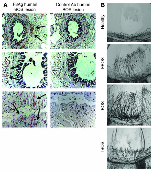
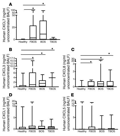
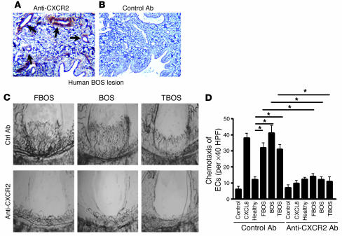
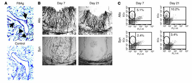
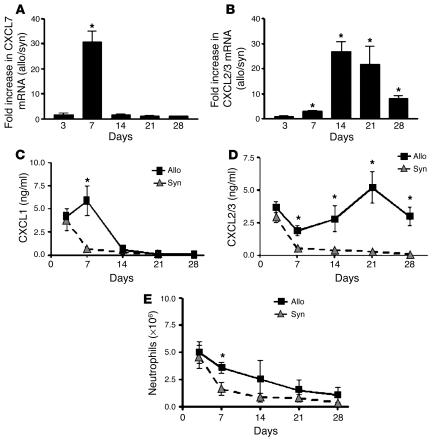
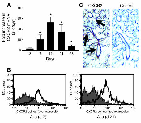
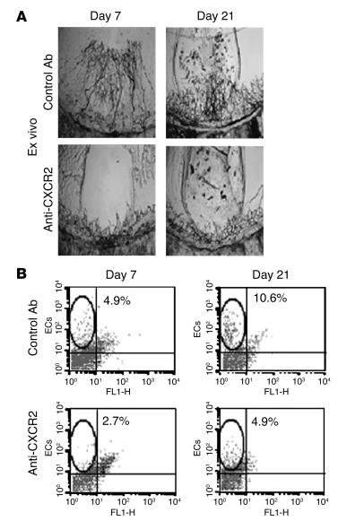
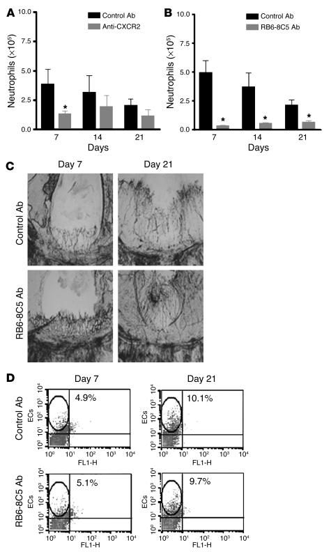
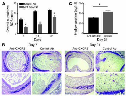
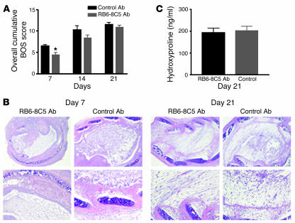
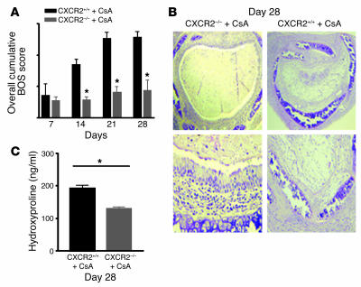
Angiogenesis and vascular remodeling support fibroproliferative processes; however, no study has addressed the importance of angiogenesis during fibro-obliteration of the allograft airway during bronchiolitis obliterans syndrome (BOS) that occurs after lung transplantation. The ELR(+) CXC chemokines both mediate neutrophil recruitment and promote angiogenesis. Their shared endothelial cell receptor is the G-coupled protein receptor CXC chemokine receptor 2 (CXCR2). We found that elevated levels of multiple ELR(+) CXC chemokines correlated with the presence of BOS. Proof-of-concept studies using a murine model of BOS not only demonstrated an early neutrophil infiltration but also marked vascular remodeling in the tracheal allografts. In addition, tracheal allograft ELR(+) CXC chemokines were persistently expressed even in the absence of significant neutrophil infiltration and were temporally associated with vascular remodeling during fibro-obliteration of the tracheal allograft. Furthermore, in neutralizing studies, treatment with anti-CXCR2 Abs inhibited early neutrophil infiltration and later vascular remodeling, which resulted in the attenuation of murine BOS. A more profound attenuation of fibro-obliteration was seen when CXCR2(-/-) mice received cyclosporin A. This supports the notion that the CXCR2/CXCR2 ligand biological axis has a bimodal function during the course of BOS: early, it is important for neutrophil recruitment and later, during fibro-obliteration, it is important for vascular remodeling independent of neutrophil recruitment.


Figures

Figure 1
Figure 1
Vascular remodeling occurs during the pathogenesis of human BOS. (A) Representative photomicrographs of the immunolocalization of F8Ag in BOS lung tissue. Magnification, ×40 (top row); ×100 (middle row); and ×200 (bottom row). (B) Representative photographs of corneal vascular remodeling in response to BALF specimens from healthy lung transplant recipients and recipients with FBOS, BOS, and TBOS. Magnification, ×20.
Figure 2
Figure 2
Human CXCL7 (A), CXCL3 (B), CXCL8 (C), CXCL1 (D), and CXCL5 (E) protein levels in unconcentrated BALF from recipients with FBOS, BOS, and TBOS and healthy lung transplant recipients in a 4-group comparison. *P < 0.001.
Figure 3
Figure 3
Representative photomicrographs of the immunolocalization of CXCR2 in human BOS lung tissue. BOS lung specimen immunostained for CXCR2 demonstrating immunolocalization to the vascular endothelium (A) and BOS lung specimen immunostained with control Ab (B). Magnification, ×400. (C) Representative photomicrograph of corneal vascular remodeling in response to FBOS, BOS, and TBOS BALF specimens with neutralizing anti-CXCR2 Ab compared with control Ab. Magnification, ×20. (D) BALF ELR+ CXC chemokines from patients with FBOS, BOS, and TBOS are biologically angiogenic as determined by HLMVEC chemotaxis. There is significantly more chemotaxis to the FBOS, BOS, and TBOS BALF compared with that from healthy transplant recipients. Neutralizing Abs to CXCR2 inhibited chemotaxis in the FBOS, BOS and TBOS groups, but had no effect on the healthy group. *P < 0.05.
Figure 4
Figure 4
Vascular remodeling occurs during the pathogenesis of murine BOS. (A) Representative photomicrographs of immunolocalization of F8Ag in a murine fibro-obliteration lesion at day 21 as compared with control Ab. Magnification, ×400. (B) CMP assay for vascular remodeling of tracheal-derived angiogenic activity from allografts (Allo) and syngeneic controls (Syn) at days 7 and 21. Magnification, ×20. (C) FACS analysis of MECA-32 from single-cell suspension of allograft and syngeneic control digests at days 7 and 21.
Figure 5
Figure 5
ELR+ CXC chemokines are elevated during the pathogenesis of murine BOS. Real-time quantitative PCR of CXCL1 (A) and CXCL2/3 (B) mRNA expression presented as fold increase in chemokine expression in allografts compared with syngeneic controls at days 3, 7, 14, 21, and 28. Protein levels of CXCL1 (C) and CXCL2/3 (D) in allografts and syngeneic controls at days 3 through 28. (E) FACS analysis of neutrophils from tracheal allografts undergoing BOS as compared with syngeneic controls. *P < 0.05.
Figure 6
Figure 6
Endothelial cells have a marked increase in CXCR2 expression during the pathogenesis of murine BOS. (A) Real-time quantitative PCR determination of CXCR2 mRNA expression presented as fold increase in chemokine expression in allografts compared with syngeneic controls at days 3, 7, 14, 21, and 28. *P < 0.05. (B) FACS analysis of gated MECA-32 cells from tracheal allografts for cell surface protein expression of CXCR2 at days 7 and 21. (C) Representative photomicrographs of immunolocalization of CXCR2 in a murine fibro-obliteration lesion at day 21 compared with control Abs. Magnification, ×400.
Figure 7
Figure 7
Vascular remodeling and angiogenic activity during murine BOS is attributable to CXCR2/CXCR2 ligand biology. (A) Representative CMP assay for vascular remodeling of tracheal-derived angiogenic activity from allografts with ex vivo–administered anti-CXCR2 Ab compared with control Ab. Magnification, ×20. (B) FACS analysis of MECA-32 from allografts treated with in vivo anti-CXCR2 Ab compared with control Ab.
Figure 8
Figure 8
Inhibition of CXCR2/CXCR2 ligand biology attenuates vascular remodeling and angiogenic activity independent of its effects on neutrophils during murine BOS. (A) FACS analysis of neutrophils in tracheal allografts from recipients treated with anti-CXCR2 compared with control Ab. (B) FACS analysis of neutrophils in tracheal allografts from recipients treated with RB6-8C5 Ab compared with control Ab. *P < 0.05. (C) Representative CMP assay for vascular remodeling of tracheal-derived angiogenic activity from allografts with in vivo–administered RB6-8C5 Ab compared with control Ab at days 7 and 21. Magnification, ×20. (D) FACS analysis of MECA-32 from allografts treated in vivo with RB6-8C5 Ab compared with control Ab.
Figure 9
Figure 9
Inhibition of CXCR2/CXCR2 ligand biology attenuates murine BOS. (A) Quantitative analysis of histopathologic sections of tracheal allografts from animals treated with anti-CXCR2 Ab compared with control Ab. (B) Representative photomicrographs of the histopathology of tracheal allografts from animals treated with anti-CXCR2 Ab compared with control Ab at days 7 and 21. Magnification, ×40 (top row) and ×100 (bottom row). (C) Hydroxyproline levels in tracheal allografts from animals treated with anti-CXCR2 Ab compared with control Ab at day 21. *P < 0.05.
Figure 10
Figure 10
Neutrophil depletion does not affect late fibro-obliteration during the pathogenesis of murine BOS. (A) Quantitative analysis of histopathologic sections of tracheal allografts from animals treated with RB6-8C5 Ab compared with control Ab. (B) Representative photomicrographs of the histopathology of tracheal allografts from animals treated with RB6-8C5 Ab compared with control Ab at days 7 and 21. Magnification, ×40 (top row) and ×100 (bottom row). (C) Hydroxyproline levels in tracheal allografts from animals treated with RB6-8C5 Ab compared with control Ab at day 21. *P < 0.05.
Figure 11
Figure 11
Neutrophil depletion plus anti-CXCR2 Ab treatment has no synergistic or additive effect on murine BOS as compared to anti-CXCR2 treatment alone. (A) Quantitative analysis of histopathologic sections of tracheal allografts from animals treated with: anti-CXCR2 Ab plus control 1 Ab (CTRL1); anti-CXCR2 Ab plus RB6-8C5 Ab; RB6-8C5 Ab plus control 2 Ab (CTRL2); and control 1 Ab plus control 2 Ab. (B) Hydroxyproline levels in tracheal allografts from animals treated with: anti-CXCR2 Ab plus control 1 Ab; anti-CXCR2 Ab plus RB6-8C5 Ab; RB6-8C5 Ab plus control 2 Ab; and control 1 Ab plus control 2 Ab. *P < 0.0083.
Figure 12
Figure 12
CXCR2–/– recipients of tracheal allografts treated with CsA attenuates murine BOS for a prolonged period of time. (A) Quantitative analysis of histopathologic sections of tracheal allografts from CXCR2–/– animals treated with CsA compared with CXCR2+/+ animals treated with CsA. (B) Representative photomicrographs of the histopathology of tracheal allografts from CXCR2–/– animals treated with CsA compared with CXCR2+/+ animals treated with CsA at day 28, Magnification, ×40 (top row) and ×100 (bottom row). (C) Hydroxyproline levels in tracheal allografts from CXCR2–/– animals treated with CsA compared with CXCR2+/+ animals treated with CsA at day 28. *P < 0.05.

Comment in

References

    1. Trulock EP. Lung transplantation. Am. J. Respir. Crit. Care Med. 1997;155:789–818. - PubMed
    1. Arcasoy SM, Kotloff RM. Lung transplantation. N. Engl. J. Med. 1999;340:1081–1091. - PubMed
    1. Folkman J. Angiogenesis in cancer, vascular, rheumatoid and other disease. Nat. Med. 1995;1:27–31. - PubMed
    1. Belperio JA, et al. CXC chemokines in angiogenesis. J. Leukoc. Biol. 2000;68:1–8. - PubMed
    1. Strieter RM, Belperio JA, Keane MP. CXC chemokines in vascular remodeling related to pulmonary fibrosis. Am. J. Respir. Cell Mol. Biol. 2003;29(Suppl. 3):S67–S69. - PubMed

Publication types