. 2005 Aug 15;390(Pt 1):303-9.
doi: 10.1042/BJ20050375.

Enhanced laminin binding by alpha-dystroglycan after enzymatic deglycosylation

Affiliations

Enhanced laminin binding by alpha-dystroglycan after enzymatic deglycosylation

Ariana C Combs et al. Biochem J. .

Abstract

Carbohydrate modifications are clearly important to the function of alpha-dystroglycan but their composition and structure remain poorly understood. In the present study, we describe experiments aimed at identifying the alpha-dystroglycan oligosaccharides important for its binding to laminin-1 and carbohydrate-dependent mAbs (monoclonal antibodies) IIH6 and VIA4(1). We digested highly purified skeletal muscle alpha-dystroglycan with an array of linkage-specific endo- and exoglycosidases, which were verified for action on alpha-dystroglycan by loss/gain of reactivity for lectins with defined glyco-epitopes. Notably, digestion with a combination of Arthrobacter ureafaciens sialidase, beta(1-4)galactosidase and beta-N-acetylglucosaminidase substantially degraded SiaAalpha2-3Galbeta1-4GlcNAcbeta1-2Man glycans on highly purified alpha-dystroglycan that nonetheless exhibited enhanced IIH6, VIA4(1) and laminin-1 binding activity. Additional results indicate that alpha-dystroglycan is probably modified with other anionic sugars besides sialic acid and suggest that rare alpha-linked GlcNAc moieties may block its complete deglycosylation with currently available enzymes.


Figures

Figure 1
Figure 1. Characterization of purified skeletal muscle α-dystroglycan
Shown in (A) are a Coomassie Blue-stained gel (CB) and an autoradiograph from a gel loaded with iodinated α-dystroglycan (125I-αDG). Also shown are nitrocellulose transfers loaded with purified α-dystroglycan and blotted with polyclonal (αDG pAb) or mAb (IIH6) to α-dystroglycan, ConA, or laminin-1. Molecular mass standards (×10−3) are indicated on the left. Shown in (B) is the amount of 125I-α-dystroglycan specifically bound to microtitre wells coated with laminin-1 as a function of increasing α-dystroglycan concentration. Shown in (C) is the amount of laminin-1 bound to microtitre wells coated with purified α-dystroglycan in the presence of increasing concentrations of heparin or N-acetylneuraminic acid (NANA).
Figure 2
Figure 2. Heterogeneous sialoglycosylation of skeletal muscle α-dystroglycan
Shown are identical nitrocellulose transfers loaded with equal amounts of purified α-dystroglycan incubated for 17 h at 37 °C in 50 mM sodium phosphate (pH 7.0) in the absence (Con), or presence of A. ureafaciens, C. perfringens or V. cholerae sialidase and stained with α-dystroglycan mAb IIH6, horseradish peroxidase-conjugated WGA or LFA.
Figure 3
Figure 3. Enzymatic deglycosylation of skeletal muscle α-dystroglycan
α-Dystroglycan was incubated for 20 h at 37 °C in 50 mM sodium phosphate, pH 7, in the absence (−) or presence (+) of all five enzymes provided in the GLYCOPRO (N-glycosidase F, A. ureafaciens sialidase, and endo-O-glycosidase) and PROLINK Extender [β(1-4)galactosidase and β-N-acetylglucosaminidase] enzymatic deglycosylation kits. In (A), equal amounts of control (−) and GLYCOPRO-treated (+) α-dystroglycan were resolved on SDS-polyacrylamide gels and stained with Coomassie Blue, or transferred on to nitrocellulose and stained with chicken polyclonal antibodies to α-dystroglycan core protein (α-DG pAb). Identical gels/transfers containing equal amounts of control and enzyme-treated α-dystroglycan were analysed for residual carbohydrate (Dig-Hz), Stains-All reactivity, or binding to the lectins LFA or ConA. The asterisk marks the 180000 Mr endo-O-glycosidase band present in the enzyme-treated samples. In (B), identically prepared transfers were analysed for laminin-1 binding (Ln-1 O/L), or for reactivity with mAbs IIH6 and VIA41.
Figure 4
Figure 4. Skeletal muscle α-dystroglycan contains latent glyco-epitopes reactive with conA and GsII
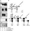
Shown in (A) are identical nitrocellulose transfers loaded with equal amounts of purified α-dystroglycan incubated for 18 h at 37 °C in 50 mM sodium phosphate (pH 7) in the absence or presence of the indicated enzymes and stained with α-dystroglycan mAb IIH6, PNA, ECA, GsII lectin or ConA. Shown in (B) are the sialylated core 1 (top) and O-mannosyl (bottom) oligosaccharide structures known to modify α-dystroglycan with predicted effect of each glycosidase on oligosaccharide structure and lectin reactivity.
Figure 5
Figure 5. Periodate oxidation of α-dystroglycan
Shown are identical nitrocellulose transfers loaded with equal amounts of skeletal muscle α-dystroglycan incubated at room temperature in 10 mM sodium metaperiodate for the indicated times and analysed for laminin-1 binding (Ln-1 O/L), or for reactivity with chicken polyclonal antibodies to α-dystroglycan (α-DG pAb), mAb IIH6 or ConA.

References

    1. Blake D. J., Weir A., Newey S. E., Davies K. E. Function and genetics of dystrophin and dystrophin-related proteins in muscle. Physiol. Rev. 2002;82:291–329. - PubMed
    1. Ervasti J. M. Costameres: the Achilles' heel of Herculean muscle. J. Biol. Chem. 2003;278:13591–13594. - PubMed
    1. Michele D. E., Campbell K. P. Dystrophin–glycoprotein complex: post-translational processing and dystroglycan function. J. Biol. Chem. 2003;278:15457–15460. - PubMed
    1. Cote P. D., Moukhles H., Lindenbaum M., Carbonetto S. Chimaeric mice deficient in dystroglycans develop muscular dystrophy and have disrupted myoneural synapses. Nat. Genet. 1999;23:338–342. - PubMed
    1. Cohn R. D., Henry M. D., Michele D. E., Barresi R., Saito F., Moore S. A., Flanagan J. D., Skwarchuk M. W., Robbins M. E., Mendell J. R., et al. Disruption of DAG1 in differentiated skeletal muscle reveals a role for dystroglycan in muscle regeneration. Cell (Cambridge, Mass.) 2002;110:639–648. - PubMed

Publication types