Comparative Study
. 2005 May 2;201(9):1397-405.
doi: 10.1084/jem.20042224.

Transplantation of a multipotent cell population from human adipose tissue induces dystrophin expression in the immunocompetent mdx mouse

Affiliations
Comparative Study

Transplantation of a multipotent cell population from human adipose tissue induces dystrophin expression in the immunocompetent mdx mouse

Anne-Marie Rodriguez et al. J Exp Med. .

Abstract

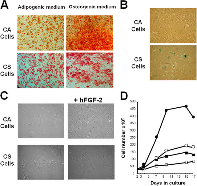
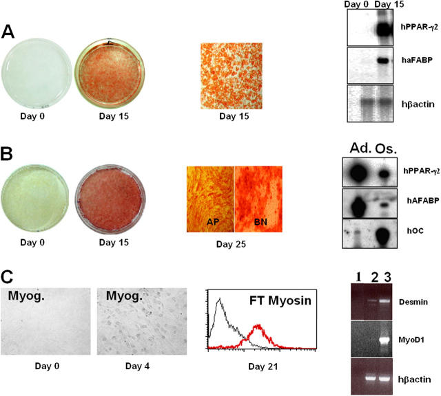
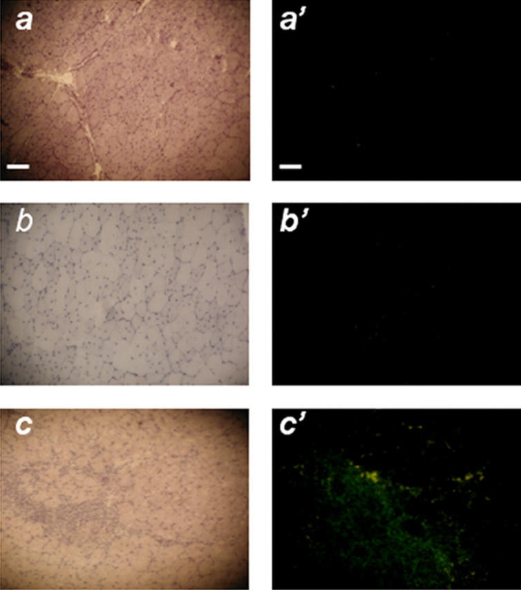
Here, we report the isolation of a human multipotent adipose-derived stem (hMADS) cell population from adipose tissue of young donors. hMADS cells display normal karyotype; have active telomerase; proliferate >200 population doublings; and differentiate into adipocytes, osteoblasts, and myoblasts. Flow cytometry analysis indicates that hMADS cells are CD44+, CD49b+, CD105+, CD90+, CD13+, Stro-1(-), CD34-, CD15-, CD117-, Flk-1(-), gly-A(-), CD133-, HLA-DR(-), and HLA-I(low). Transplantation of hMADS cells into the mdx mouse, an animal model of Duchenne muscular dystrophy, results in substantial expression of human dystrophin in the injected tibialis anterior and the adjacent gastrocnemius muscle. Long-term engraftment of hMADS cells takes place in nonimmunocompromised animals. Based on the small amounts of an easily available tissue source, their strong capacity for expansion ex vivo, their multipotent differentiation, and their immune-privileged behavior, our results suggest that hMADS cells will be an important tool for muscle cell-mediated therapy.


Figures

Figure 1.
Figure 1.
Isolation of cell populations and establishment of hMADS-2 cells. (A) In vitro differentiation of CA and CS cells at early passages (10 PDs). Cultures were stained after 15 d with Oil-red O for adipocytes and with Alizarin red for osteoblasts. (B) Cell morphology and SA β-galactosidase activity of CA and CS cells after 60 PDs. (C) Cell morphology and (D) proliferative response of CA (○, •) and CS (□, ▪) cells in the absence (○, □) or the presence of 5 ng/ml hFGF-2 (•, ▪).
Figure 2.
Figure 2.
Characteristics of culture-expanded hMADS cells. (A) A representative result of cytogenetic analysis of hMADS-2 cells (n = 3). (B) hMADS-2 cells cultured for 40 (early) or 160 (late) PDs (n = 4) were labeled with FITC-conjugated antibodies against STRO-1, CD44, CD34, CD49b, CD15, CD105, and with PE-conjugated antibodies against CD90, CD117, CD13, Flk-1, Gly-A, CD133, class I-HLA, and HLA-DR or immunoglobulin isotype control antibodies (n = 4). Black line, control immunoglobulins; red line, specific antibodies.
Figure 3.
Figure 3.
In vitro differentiation of hMADS cells (80–120 PDs) to adipocytes, osteoblasts, and myoblasts. (A) Adipogenic differentiation. Confluent hMADS-2 cells before (day 0) and 15 d after treatment with adipogenic medium were stained with Oil red O for lipid droplets. RNAs were prepared and analyzed by RT-PCR for expression of PPARγ2 and aFABP. (B) Osteogenic differentiation. Confluent hMADS-2 cells before (day 0) and 15 d after treatment with osteogenic medium were stained with Alizarin red for bone nodules. At day 25 after osteogenic induction, cells were stained for alkaline phosphatase (AP) or with Alizarin red for bone nodules (BN). RNAs were prepared and analyzed by RT-PCR for expression of PPARγ2 and aFABP (adipocyte-specific genes) and osteocalcin (OC) (osteoblast-specific gene) in adipogenic (Ad.) and osteogenic (Os.) conditions. (C) Myogenic differentiation. Confluent hMADS-2 cells before (day 0) and 4 d after treatment with myogenic medium were stained for myogenin (Myog.). Fast-twitch myosin of permeabilized cells was detected by FACS 21 d after treatment with myogenic medium. RNAs were prepared and analyzed by RT-PCR for expression of MyoD1 and desmin. (1) No RNA; (2) RNAs from undifferentiated hMADS cells; (3) RNAs from hMADS cells in myogenic medium for 4 d.
Figure 4.
Figure 4.
Human dystrophin and hMADS cells in skeletal muscle of mdx mice. (A) Expression of human dystrophin and detection of human nuclei 10 d (a–c), 50 d (d–f), 80 d (g and h), and 180 d (i) after transplantation of hMADS-2 cells. Human nuclei (nuclei counterstained with DAPI in blue and human centromeres as red hybridization signals) were found present within dystrophin-positive myofibers (green) and also at the periphery of myofibers using dystrophin antibody NCL-DYS2. (a, a′, d, d′) Right tibialis anterior muscle (control); (b, b′) left tibialis anterior muscle of cyclosporin A–treated mdx mice; (c, c′, e, e′) left tibialis anterior muscle of immunocompetent mdx mice; (f, f′), left gastrocnemius muscle of immunocompetent mdx mice; g, Left tibialis anterior muscle; h, left gastrocnemius; i, left tibialis anterior muscle. Bar, 20 μm (a–c, d–f, g–i) and 5 μm (a′–c′, d′–f′). (B) Dystrophin-positive myofibers and dystrophin subcellular localization were analyzed in the tibialis anterior muscle 10 d after transplantation using antibodies directed against (c, e) NH2 terminus of human dystrophin and (d, e) mouse collagen III. Bars, 1 μm.
Figure 5.
Figure 5.
Absence of infiltration by mCD3-positive lymphocytes in muscle of immunocompetent mdx mice after transplantation with hMADS cells. Staining was performed with hematoxylin (a–c) or with antibodies against mouse CD3 (a′, c′); left tibialis anterior muscle nontransplanted (a, a′) or 10 d after transplantation of hMADS-2 cells (b, b′) (n = 8) and CA cells at 40 PDs (c, c′) (n = 5). Bar, 50 μm (a–c) and 20 μm (a′–c′).

References

    1. Verfaillie, C.M. 2002. Adult stem cells: assessing the case for pluripotency. Trends Cell Biol. 12:502–508. - PubMed
    1. Pittenger, M.F., A.M. Mackay, S.C. Beck, R.K. Jaiswal, R. Douglas, J.D. Mosca, M.A. Moorman, D.W. Simonetti, S. Craig, and D.R. Marshak. 1999. Multilineage potential of adult human mesenchymal stem cells. Science. 284:143–147. - PubMed
    1. Krause, D.S., N.D. Theise, M.I. Collector, O. Henegariu, S. Hwang, R. Gardner, S. Neutzel, and S.J. Sharkis. 2001. Multi-organ, multi-lineage engraftment by a single bone marrow-derived stem cell. Cell. 105:369–377. - PubMed
    1. Jiang, Y., B. Vaessen, T. Lenvik, M. Blackstad, M. Reyes, and C.M. Verfaillie. 2002. Multipotent progenitor cells can be isolated from postnatal murine bone marrow, muscle and brain. Exp. Hematol. 30:896–904. - PubMed
    1. Jiang, Y., B.N. Jahagirdar, R.L. Reinhardt, R.E. Schwartz, C.D. Keene, X.R. Ortiz-Gonzalez, M. Reyes, T. Lenvik, T. Lund, M. Blackstad, et al. 2002. Pluripotency of mesenchymal stem cells derived from adult marrow. Nature. 118:41–49. - PubMed

Publication types

MeSH terms