

Forced expression of alpha-myosin heavy chain in the rabbit ventricle results in cardioprotection under cardiomyopathic conditions
- PMID: 15867177
- PMCID: PMC1314981
- DOI: 10.1161/01.CIR.0000164233.09448.B1
Forced expression of alpha-myosin heavy chain in the rabbit ventricle results in cardioprotection under cardiomyopathic conditions
Abstract
Background: The biochemical differences between the 2 mammalian cardiac myosin heavy chains (MHCs), alpha-MHC and beta-MHC, are well described, but the physiological consequences of basal isoform expression and isoform shifts in response to altered cardiac load are not clearly understood. Mature human ventricle contains primarily the beta-MHC isoform. However, the alpha-MHC isoform can be detected in healthy human ventricle and appears to be significantly downregulated in failing hearts. The unique biochemical properties of the alpha-MHC isoform might offer functional advantages in a failing heart that is expressing only the beta-MHC isoform. This hypothesis cannot be tested in mice or rats because both species express alpha-MHC as the predominant isoform.
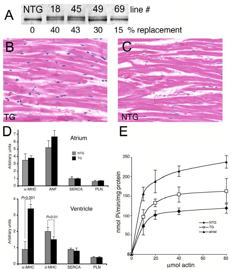
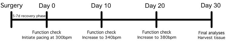
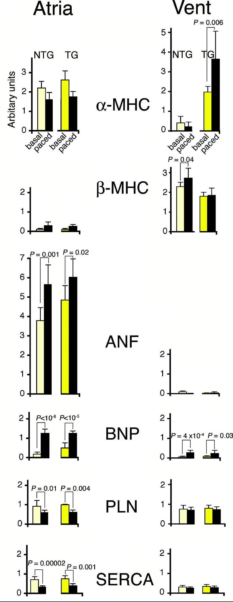
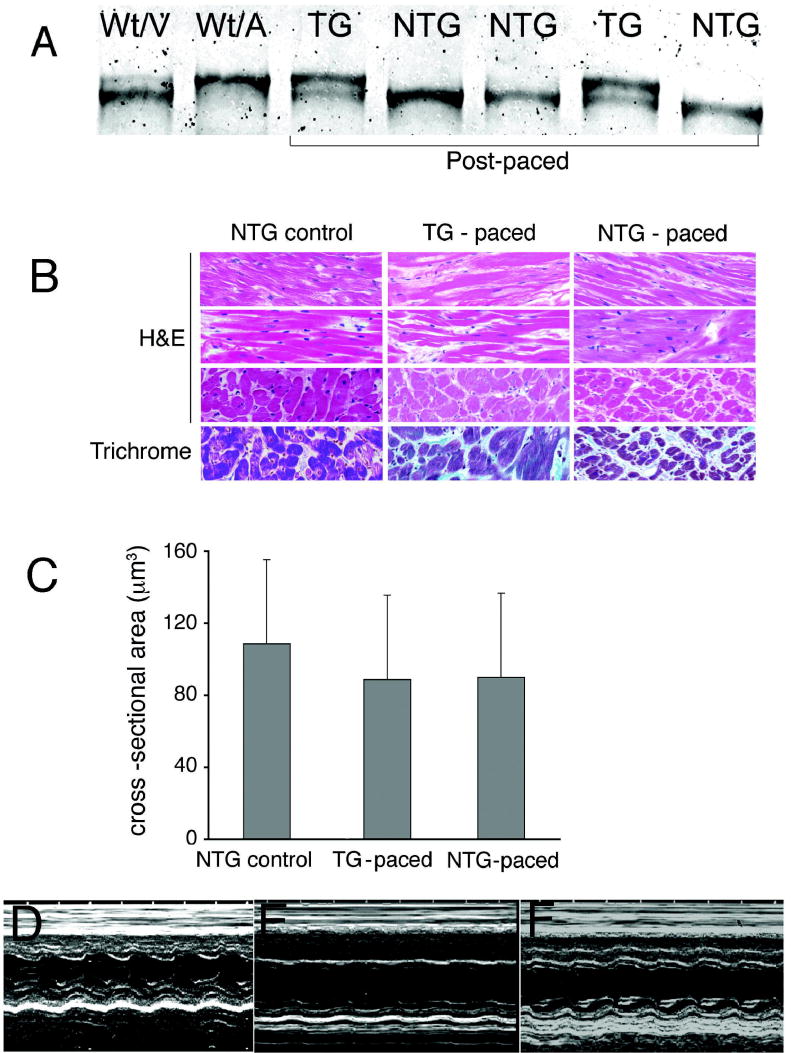
Methods and results: To test the effects of persistent alpha-MHC expression on the background of beta-MHC, we made transgenic (TG) rabbits that expressed rabbit alpha-MHC cDNA in the ventricle so that the endogenous myosin was partially replaced by the transgenically encoded species. Molecular, histological, and functional analyses showed no significant baseline effects in the TG rabbits compared with nontransgenic (NTG) littermates. To determine whether alpha-MHC expression afforded any advantages to stressed myocardium, a cohort of TG and NTG rabbits was subjected to rapid ventricular pacing. Although both the TG and NTG rabbits developed dilated cardiomyopathy, the TG rabbits had a higher shortening fraction, less septal thinning, and more normal +/-dP/dt than paced NTG rabbits.
Conclusions: Transgenic expression of alpha-MHC does not have any apparent detrimental effects under basal conditions and is cardioprotective in experimental tachycardia-induced cardiomyopathy.
Figures
Comment in
-
On mice, rabbits, and human heart failure.Circulation. 2005 May 10;111(18):2276-9. doi: 10.1161/01.CIR.0000167559.13502.9A. Circulation. 2005. PMID: 15883223 Free PMC article.
References
-
- Lowes BD, Minobe W, Abraham WT, Rizeq MN, Bohlmeyer TJ, Quaife RA, Roden RL, Dutcher DL, Robertson AD, Voelkel NF, Badesch DB, Groves BM, Gilbert EM, Bristow MR. Changes in gene expression in the intact human heart. Downregulation of alpha-myosin heavy chain in hypertrophied, failing ventricular myocardium. J Clin Invest. 1997;100:2315–2324. - PMC - PubMed
-
- Miyata S, Minobe W, Bristow MR, Leinwand LA. Myosin heavy chain isoform expression in the failing and nonfailing human heart. Circ Res. 2000;86:386–390. - PubMed
-
- Reiser PJ, Portman MA, Ning XH, Schomisch Moravec C. Human cardiac myosin heavy chain isoforms in fetal and failing adult atria and ventricles. Am J Physiol Heart Circ Physiol. 2001;280:H1814–1820. - PubMed
-
- Abraham WT, Gilbert EM, Lowes BD, Minobe WA, Larrabee P, Roden RL, Dutcher D, Sederberg J, Lindenfeld JA, Wolfel EE, Shakar SF, Ferguson D, Volkman K, Linseman JV, Quaife RA, Robertson AD, Bristow MR. Coordinate changes in Myosin heavy chain isoform gene expression are selectively associated with alterations in dilated cardiomyopathy phenotype. Mol Med. 2002;8:750–760. - PMC - PubMed
Publication types
MeSH terms
Substances
Grants and funding
LinkOut - more resources
Full Text Sources
Other Literature Sources
Research Materials
Miscellaneous
