Review
. 2005:34:267-94.
doi: 10.1146/annurev.biophys.34.040204.144452.

Structural and sequence motifs of protein (histone) methylation enzymes

Affiliations
Review

Structural and sequence motifs of protein (histone) methylation enzymes

Xiaodong Cheng et al. Annu Rev Biophys Biomol Struct. 2005.

Abstract

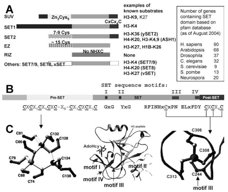
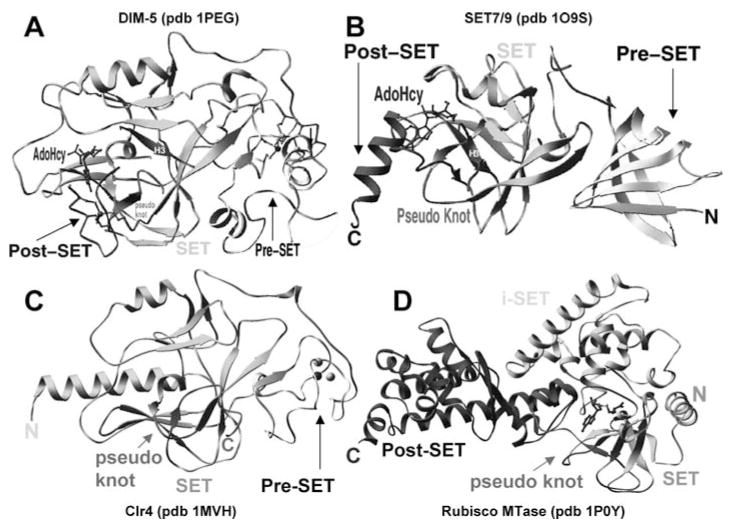
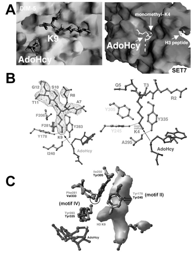
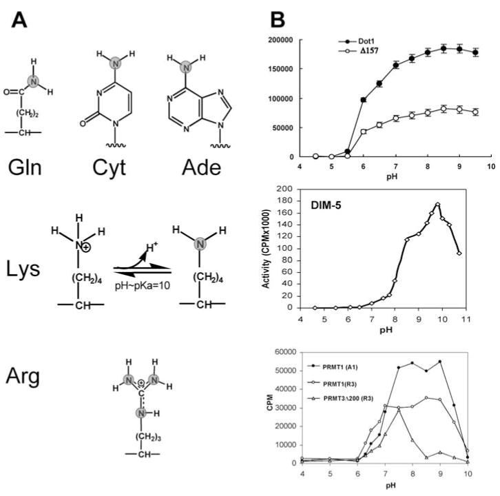
With genome sequencing nearing completion for the model organisms used in biomedical research, there is a rapidly growing appreciation that proteomics, the study of covalent modification to proteins, and transcriptional regulation will likely dominate the research headlines in the next decade. Protein methylation plays a central role in both of these fields, as several different residues (Arg, Lys, Gln) are methylated in cells and methylation plays a central role in the "histone code" that regulates chromatin structure and impacts transcription. In some cases, a single lysine can be mono-, di-, or trimethylated, with different functional consequences for each of the three forms. This review describes structural aspects of methylation of histone lysine residues by two enzyme families with entirely different structural scaffolding (the SET proteins and Dot1p) and methylation of protein arginine residues by PRMTs.


Figures

Figure 1
Figure 1
SET domain HKMTs. (a) Domain structure of SET HKMT families. (b) DIM-5 protein (one of the smallest members of the SUV family) contains four segments: a weakly conserved N-terminal region, a pre-SET domain containing nine invariant cysteines, the SET region containing four signature motifs, and the post-SET domain containing three invariant cysteines. (c) Illustration of pre-SET Zn3Cys9 triangular zinc cluster (left panel); ribbon diagram of DIM-5 SET domain, with arrows indicating locations of conserved motifs, the cofactor binding and substrate histone H3 peptide, and the pseudo knot formed by motifs III and IV (middle panel); and post-SET zinc center (right panel).
Figure 2
Figure 2
Representative examples of SET domain containing structures. (a) Neurospora DIM-5, (b) human SET7/9, (c) S. pombe Clr4, and (d) Rubisco MTase.
Figure 3
Figure 3
Active site of SET domain. (a) H3 peptide-binding site in DIM-5 with the target Lys-9 inserted into a channel (PDB 1PEG) (left panel), and the AdoHcy-binding site in SET7/9, located at the opposite end of the target lysine-binding channel (PDB 1O9S) (right panel). (b) The active sites in DIM-5 (PDB 1PEG) (left panel) and SET7/9 (PDB 1MT6) (right panel). The arrow indicates the movement of the methyl group transferred from the AdoMet methylsulfonium group to the target amino group. (c) Structural comparison of active sites in DIM-5 and SET7/9: either two tyrosines and one phenylalanine (DIM-5) or three tyrosines (SET7/9) surrounds the target lysine.
Figure 4
Figure 4
Mass spectrometry analysis of methylation products for (a) WT DIM-5 and its F281Y variant (113), (b) WT SET7/9 and its Y305F variant (113), and (c) G9a and its F1205Y variant (12a).
Figure 5
Figure 5
Dot1p family (non-SETHKMTs). (a) Schematic representation of Dot1 homologues from yeast and human. (b) Dot1 core structure: (left panel) human Dot1L (residues 5–332) in complex with methyl-donor AdoMet (PDB1NW3) and (right panel) yeast Dot1p (residues 176–567) incomplex with reaction by-product AdoHcy (PDB1U2Z). The N-terminal helical domain and the C-terminal catalytic domain are circled. The bound methyl-donor AdoMet in human Dot1L and there action by-product AdoHcy are shown as stick models. The largest conformational difference (indicated by arrows) between the two catalytic domains is the hairpin loop-containing motif VIII forming part of the active site. (c) Five hydrophobic residues of yeast Dot1p form the active-site pocket (the corresponding residue from human Dot1L is in parenthesis) (left panel). The opening of the pocket is approximately 4×5 Å, into which the target lysine could be inserted (middle panel). A surface representation of yeast Dot1p core showing the conserved motifs (X, VI, IV, and VIII) surrounding the active-site pocket, through which only the AdoHcy sulfur atom is visible (right panel). Conserved motifs (I, II, and III) involved in interacting with AdoHcy are buried and invisible from the surface.
Figure 6
Figure 6
(a) Examples of known targets of amino methylation. Only the deprotonated amino group (NH2) has a free lone pair of electrons capable of nucleophilic attack on the AdoMet methyl group. (b) Methyl transfer activities (measured as TCA precipitable counts) as function of pH: (top panel) yeast Dot1p and its N-terminal deletion mutant Δ 157 (77), (middle panel) DIM-5 (112), and (bottom panel) rat PRMT1, rat PRMT3 (full length), and its PRMT core domain (Δ 200) (111).
Figure 7
Figure 7
(a) Two major types of protein arginine methylation. (b) Members of PRMT family. The conserved MTase domain is in black and the unique β-barrel domain to the PRMT family is in gray. The N and C termini of the proteins and the first invariant residue are labeled. (c) Dimer structures of PRMT cores: (left panel) rat PRMT1, (middle panel) rat PRMT3, and (right panel) yeast RMT1/Hmt1.
Figure 8
Figure 8
Peptide-binding grooves (P1, P2, and P3) in the structure of ternary complex of PRMT1-AdoHcy-R3 peptide (sequence shown at the bottom): solvent-accessible molecular surface with bound AdoHcy and arginine shown as stick models and indicated by the arrows (left panel). If the central Arg-9 were the target bound in the active site, connecting peptide-binding sites P1 and P2 would cover the active site and the entire length of the peptide. When the end arginine (either Arg-3 or Arg-15) is bound in the active site, connection of peptide-binding sites P2 and P3 would account for the length of the whole peptide (right panel). Site P3 corresponds to one of the grooves perpendicular to the strands of the β-barrel domain.

References

    1. Abramovich C, Yakobson B, Chebath J, Revel M. A protein-arginine methyltransferase binds to the intracytoplasmic domain of the IFNAR1 chain in the type I interferon receptor. EMBO J. 1997;16:260–66. - PMC - PubMed
    1. An W, Kim J, Roeder RG. Ordered cooperative functions of PRMT1, p300, and CARM1 in transcriptional activation by p53. Cell. 2004;117:735–48. - PubMed
    1. Bauer UM, Daujat S, Nielsen SJ, Nightingale K, Kouzarides T. Methylation at arginine 17 of histone H3 is linked to gene activation. EMBO Rep. 2002;3:39–44. - PMC - PubMed
    1. Baumbusch LO, Thorstensen T, Krauss V, Fischer A, Naumann K, et al. The Arabidopsis thaliana genome contains at least 29 active genes encoding SET domain proteins that can be assigned to four evolutionarily conserved classes. Nucleic Acids Res. 2001;29:4319–33. - PMC - PubMed
    1. Bode W, Fernandez-Catalan C, Tschesche H, Grams F, Nagase H, Maskos K. Structural properties of matrix metalloproteinases. Cell Mol Life Sci. 1999;55:639–52. - PMC - PubMed

Publication types

MeSH terms

LinkOut - more resources