

Post-translationally modified residues of native human osteopontin are located in clusters: identification of 36 phosphorylation and five O-glycosylation sites and their biological implications
- PMID: 15869464
- PMCID: PMC1184582
- DOI: 10.1042/BJ20050341
Post-translationally modified residues of native human osteopontin are located in clusters: identification of 36 phosphorylation and five O-glycosylation sites and their biological implications
Abstract
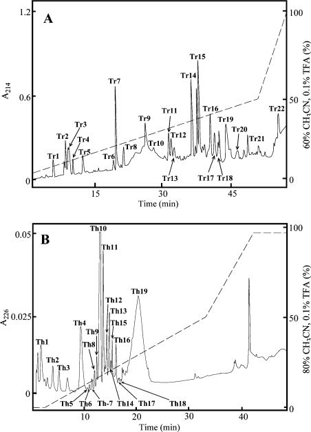
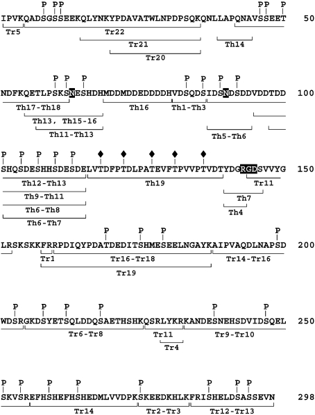
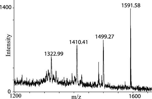
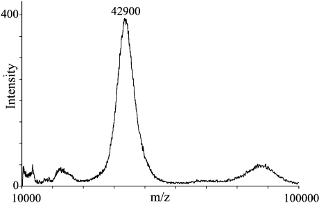
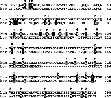
OPN (osteopontin) is an integrin-binding highly phosphorylated glycoprotein, recognized as a key molecule in a multitude of biological processes such as bone mineralization, cancer metastasis, cell-mediated immune response, inflammation and cell survival. A significant regulation of OPN function is mediated through PTM (post-translational modification). Using a combination of Edman degradation and MS analyses, we have characterized the complete phosphorylation and glycosylation pattern of native human OPN. A total of 36 phosphoresidues have been localized in the sequence of OPN. There are 29 phosphorylations (Ser8, Ser10, Ser11, Ser46, Ser47, Thr50, Ser60, Ser62, Ser65, Ser83, Ser86, Ser89, Ser92, Ser104, Ser110, Ser113, Thr169, Ser179, Ser208, Ser218, Ser238, Ser247, Ser254, Ser259, Ser264, Ser275, Ser287, Ser292 and Ser294) located in the target sequence of MGCK (mammary gland casein kinase) also known as the Golgi kinase (S/T-X-E/S(P)/D). Six phosphorylations (Ser101, Ser107, Ser175, Ser199, Ser212 and Ser251) are located in the target sequence of CKII (casein kinase II) [S-X-X-E/S(P)/D] and a single phosphorylation, Ser203, is not positioned in the motif of either MGCK or CKII. The 36 phosphoresidues represent the maximal degree of modification since variability at many sites was seen. Five threonine residues are O-glycosylated (Thr118, Thr122, Thr127, Thr131 and Thr136) and two potential sites for N-glycosylation (Asn63 and Asn90) are not occupied in human milk OPN. The phosphorylations are arranged in clusters of three to five phosphoresidues and the regions containing the glycosylations and the RGD (Arg-Gly-Asp) integrin-binding sequence are devoid of phosphorylations. Knowledge about the positions and nature of PTMs in OPN will allow a rational experimental design of functional studies aimed at understanding the structural and functional interdependences in diverse biological processes in which OPN is a key molecule.
Figures
References
-
- Fisher L. W., Torchia D. A., Fohr B., Young M. F., Fedarko N. S. Flexible structures of SIBLING proteins, bone sialoprotein, and osteopontin. Biochem. Biophys. Res. Commun. 2001;280:460–465. - PubMed
-
- Qin C., Baba O., Butler W. T. Post-translational modifications of SIBLING proteins and their roles in osteogenesis and dentinogenesis. Crit. Rev. Oral Biol. Med. 2004;15:126–136. - PubMed
-
- Sodek J., Ganss B., McKee M. D. Osteopontin. Crit. Rev. Oral Biol. Med. 2000;11:279–303. - PubMed
-
- Giachelli C. M., Steitz S. Osteopontin: a versatile regulator of inflammation and biomineralization. Matrix Biol. 2000;19:615–622. - PubMed
MeSH terms
Substances
LinkOut - more resources
Full Text Sources
Other Literature Sources
Molecular Biology Databases
Research Materials
Miscellaneous