

Crystal structure of levansucrase from the Gram-negative bacterium Gluconacetobacter diazotrophicus
- PMID: 15869470
- PMCID: PMC1188265
- DOI: 10.1042/BJ20050324
Crystal structure of levansucrase from the Gram-negative bacterium Gluconacetobacter diazotrophicus
Abstract
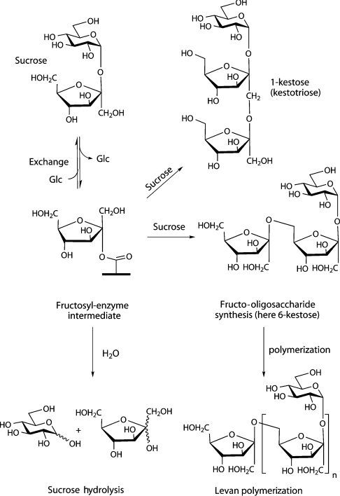
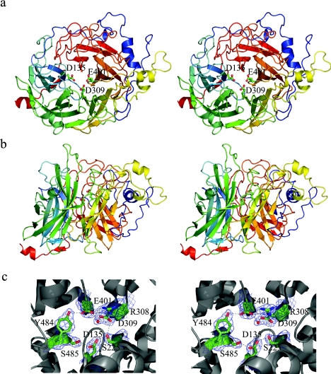
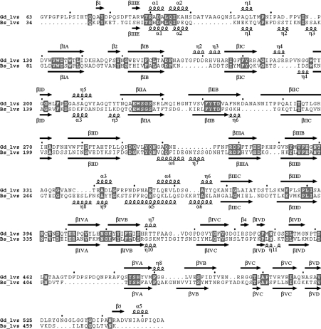
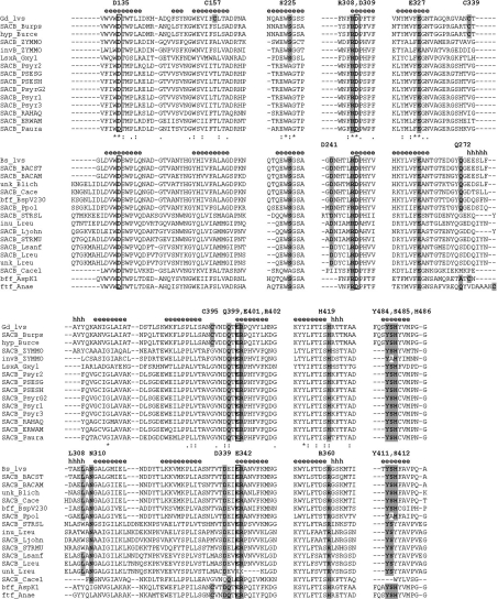
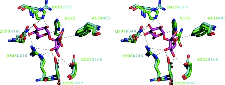
The endophytic Gram-negative bacterium Gluconacetobacter diazotrophicus SRT4 secretes a constitutively expressed levansucrase (LsdA, EC 2.4.1.10), which converts sucrose into fructooligosaccharides and levan. The enzyme is included in GH (glycoside hydrolase) family 68 of the sequence-based classification of glycosidases. The three-dimensional structure of LsdA has been determined by X-ray crystallography at a resolution of 2.5 A (1 A=0.1 nm). The structure was solved by molecular replacement using the homologous Bacillus subtilis (Bs) levansucrase (Protein Data Bank accession code 1OYG) as a search model. LsdA displays a five-bladed beta-propeller architecture, where the catalytic residues that are responsible for sucrose hydrolysis are perfectly superimposable with the equivalent residues of the Bs homologue. The comparison of both structures, the mutagenesis data and the analysis of GH68 family multiple sequences alignment show a strong conservation of the sucrose hydrolytic machinery among levansucrases and also a structural equivalence of the Bs levansucrase Ca2+-binding site to the LsdA Cys339-Cys395 disulphide bridge, suggesting similar fold-stabilizing roles. Despite the strong conservation of the sucrose-recognition site observed in LsdA, Bs levansucrase and GH32 family Thermotoga maritima invertase, structural differences appear around residues involved in the transfructosylation reaction.
Figures
Similar articles
-
The crystal structure of Erwinia amylovora levansucrase provides a snapshot of the products of sucrose hydrolysis trapped into the active site.J Struct Biol. 2015 Sep;191(3):290-8. doi: 10.1016/j.jsb.2015.07.010. Epub 2015 Jul 21. J Struct Biol. 2015. PMID: 26208466
-
A type II protein secretory pathway required for levansucrase secretion by Gluconacetobacter diazotrophicus.J Bacteriol. 2004 Aug;186(15):5031-9. doi: 10.1128/JB.186.15.5031-5039.2004. J Bacteriol. 2004. PMID: 15262940 Free PMC article.
-
Crystallization and preliminary X-ray diffraction analysis of levansucrase (LsdA) from Gluconacetobacter diazotrophicus SRT4.Acta Crystallogr D Biol Crystallogr. 2004 Jan;60(Pt 1):181-3. doi: 10.1107/s0907444903025514. Epub 2003 Dec 18. Acta Crystallogr D Biol Crystallogr. 2004. PMID: 14684923
-
[Application of levansucrase in levan synthesis--a review].Wei Sheng Wu Xue Bao. 2014 Jun 4;54(6):601-7. Wei Sheng Wu Xue Bao. 2014. PMID: 25272807 Review. Chinese.
-
A close look at the structural features and reaction conditions that modulate the synthesis of low and high molecular weight fructans by levansucrases.Carbohydr Polym. 2019 Sep 1;219:130-142. doi: 10.1016/j.carbpol.2019.05.014. Epub 2019 May 7. Carbohydr Polym. 2019. PMID: 31151510 Review.
Cited by
-
Synthesis of novel bioactive lactose-derived oligosaccharides by microbial glycoside hydrolases.Microb Biotechnol. 2014 Jul;7(4):315-31. doi: 10.1111/1751-7915.12124. Epub 2014 Apr 1. Microb Biotechnol. 2014. PMID: 24690139 Free PMC article. Review.
-
Crystal structures of Aspergillus japonicus fructosyltransferase complex with donor/acceptor substrates reveal complete subsites in the active site for catalysis.J Biol Chem. 2010 Jul 23;285(30):23251-64. doi: 10.1074/jbc.M110.113027. Epub 2010 May 13. J Biol Chem. 2010. PMID: 20466731 Free PMC article.
-
Insights into polymer versus oligosaccharide synthesis: mutagenesis and mechanistic studies of a novel levansucrase from Bacillus megaterium.Biochem J. 2007 Oct 15;407(2):189-98. doi: 10.1042/BJ20070600. Biochem J. 2007. PMID: 17608626 Free PMC article.
-
The Structure of Sucrose-Soaked Levansucrase Crystals from Erwinia tasmaniensis reveals a Binding Pocket for Levanbiose.Int J Mol Sci. 2019 Dec 20;21(1):83. doi: 10.3390/ijms21010083. Int J Mol Sci. 2019. PMID: 31877648 Free PMC article.
-
Unraveling the structural and molecular properties of 34-residue levans with various branching degrees by replica exchange molecular dynamics simulations.PLoS One. 2018 Aug 21;13(8):e0202578. doi: 10.1371/journal.pone.0202578. eCollection 2018. PLoS One. 2018. PMID: 30130368 Free PMC article.
References
-
- Cairns A. J. Fructan biosynthesis in transgenic plants. J. Exp. Bot. 2003;54:549–567. - PubMed
-
- Cote G. L., Ahlgren J. A. Metabolism in microorganisms, part I: levan and levansucrase. In: Suzuki M., Chatterton N. J., editors. Science and Technology of Fructans. Boca Raton: CRC Press; 1993. pp. 141–168.
-
- Chambert R., Treboul G., Dedonder R. Kinetic studies of levansucrase of Bacillus subtilis. Eur. J. Biochem. 1974;41:285–300. - PubMed
Publication types
MeSH terms
Substances
Associated data
- Actions
LinkOut - more resources
Full Text Sources
Other Literature Sources
Miscellaneous