Comparative Study
. 2005 Apr;21(8):2285-90.
doi: 10.1111/j.1460-9568.2005.04053.x.

Speed limits in the cerebellum: constraints from myelinated and unmyelinated parallel fibers

Affiliations
Comparative Study

Speed limits in the cerebellum: constraints from myelinated and unmyelinated parallel fibers

Krysta D Wyatt et al. Eur J Neurosci. 2005 Apr.

Abstract

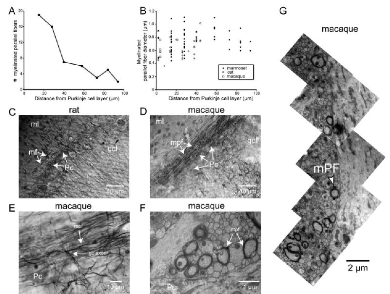
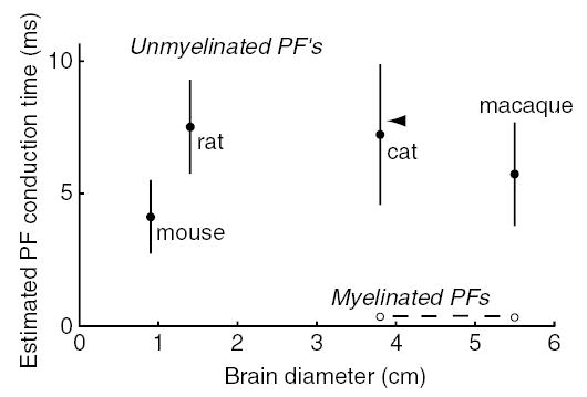
Cerebellar parallel fibers are among the thinnest known vertebrate axons and represent an extreme anatomical adaptation. Until now a systematic examination of their properties across species has not been carried out. We used transmission electron microscopy and light microscopy to compare parallel fibers in mammals of different brain sizes. From mouse to macaque, the average unmyelinated parallel fiber diameter was 0.2-0.3 microm, consistent with the idea that they are evolutionarily selected for compactness. Average unmyelinated parallel fiber diameter scaled up slightly with brain size, and across species the estimated total conduction time is 5-10 ms. However, these conduction times can vary by milliseconds, and unmyelinated PFs consume large amounts of energy per action potential. These functional disadvantages are overcome in myelinated parallel fibers, which we found in the deep regions nearest the Purkinje cell layer in marmoset, cat and macaque. These axons were 0.4-1.1 microm wide, have expected conduction times of 0.5-1.0 ms, and may convey fast feedforward inhibition via basket cells to Purkinje cells.


Figures

Fig. 1
Fig. 1
Unmyelinated and myelinated parallel fibers of the cerebellum. (A) Cerebellar anatomy (Ramon y Cajal, 1911). (B–D) Electron micrographs from marmoset cerebellum. All images are sagittal sections from lobule VI (×3150 direct magnification). Pc, Purkinje cell; Pcd, Purkinje cell dendrite; bc axon, basket cell axon; mpf, myelinated PF. (B) Lower molecular layer, adjacent to a Purkinje cell. (C) Middle molecular layer, approximately 90 μm from Purkinje cell. (D) Upper molecular layer, approximately 40 μm from the pial surface. (E) Unmyelinated PF diameters (average and SD) for five different species as a function of brain diameter. Brain diameters were obtained as described in Shultz et al. (2003), and were (in cm): mouse 0.9, rat 1.4, marmoset 2.4, cat 3.8, macaque 5.5. (F) Unmyelinated PF diameter as a function of position in molecular layer in cat. (G) Unmyelinated PF diameter as a function of distance from the Purkinje cell layer in mouse, marmoset, and macaque. In (F) and (G), each point represents data from one TEM image.
Fig. 2
Fig. 2
Myelinated parallel fibers. (A) Number of myelinated PFs as a function of position in the molecular layer. Marmoset cerebellum, lobule VI. Individual counts were taken from 78 μm2 TEM images (×3150 direct magnification, ×20 000 print magnification). (B) Myelinated PF diameter as a function of position in the molecular layer for marmoset, cat, macaque (lobule VI for all). Diameters were measured from ×20 000 and ×32 000 TEM prints (×3150 and ×5000 direct magnification). (C) Brightfield image of rat cerebellum, coronal section from crus II (×200 direct magnification), stained for myelin. Pc, Purkinje cell; gcl, granule cell layer; ml, molecular layer; mf, myelinated fiber. Note the sparse plexus of myelinated fibers in the lower molecular layer, putatively corresponding to recurrent collaterals of Purkinje cell axons and/or Lugaro cell axons. (D and E) Brightfield images of coronal sections of macaque cerebellum lobule III stained for myelin taken at (D) ×200 direct magnification and (E) ×1000 direct magnification. Pc, Purkinje cell; gcl, granule cell layer; gc axon, granule cell axon; ml, molecular layer; mpf, myelinated parallel fiber. Note the many myelinated PFs coursing through the lower molecular layer above the Purkinje cell layer and the T-junction in E. (F) TEM micrograph of macaque cerebellum, lobule VI, ×5000 direct magnification. Pc, Purkinje cell; mpf, myelinated PF. (G) Montage of multiple micrographs (×5000 direct magnification) of macaque lobule VI tissue in the lower molecular layer to show multiple myelinated PFs. The bottom of the montage begins approximately 5 μm from the top of the Purkinje cell layer.
Fig. 3
Fig. 3
Estimated parallel fiber conduction times. Estimated conduction times based on measured PF diameters corrected for shrinkage and literature values for PF lengths (mouse, Soha et al., 1997; rat, Pichitpornchai et al., 1994; cat, Brand et al., 1976; macaque, Mugnaini, 1983), halved to give an estimate of the distance from the T-junction to the end of the PF. Conduction velocities were calculated as v = 0.75 × diameter for unmyelinated axons (Hoffmeister et al., 1991), and as v = 5.7 × diameter for myelinated axons (Hursh, 1939) (diameters in μm, velocities in m/s). Arrowhead, range of auditory evoked potential latencies in Purkinje cell simple spikes in cat (Huang & Liu, 1985).

References

    1. Albus JS. A theory of cerebellar function. Math Biosci. 1971;10:25–61.
    1. Bower JM. The organization of cerebellar cortical circuitry revisited: implications for function. Ann NY Acad Sci. 2002;978:135–155. - PubMed
    1. Braitenberg V. Is the cerebellar cortex a biological clock in the millisecond range? Prog Brain Res. 1967;25:334–346. - PubMed
    1. Brand S, Dahl AL, Mugnaini E. The length of parallel fibers in the cat cerebellar cortex. An experimental light and electron microscopic study. Exp Brain Res. 1976;26:39–58. - PubMed
    1. Chadderton P, Margrie TW, Hausser M. Integration of quanta in cerebellar granule cells during sensory processing. Nature. 2004;428:856–860. - PubMed

Publication types

MeSH terms