

N-acetyl-L-cysteine ameliorates the inflammatory disease process in experimental autoimmune encephalomyelitis in Lewis rats
- PMID: 15869713
- PMCID: PMC1097751
- DOI: 10.1186/1740-2557-2-4
N-acetyl-L-cysteine ameliorates the inflammatory disease process in experimental autoimmune encephalomyelitis in Lewis rats
Abstract
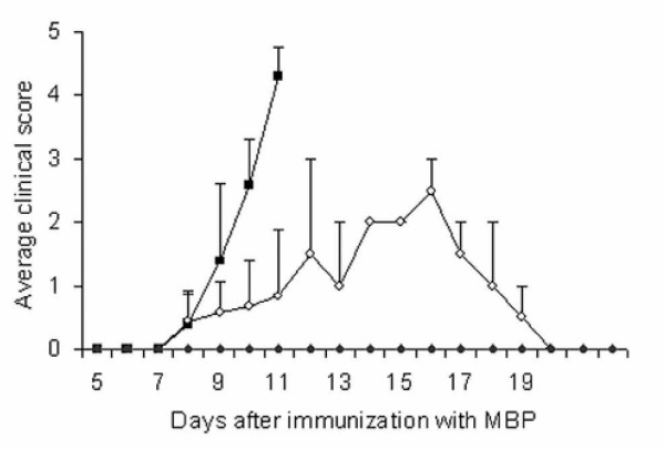
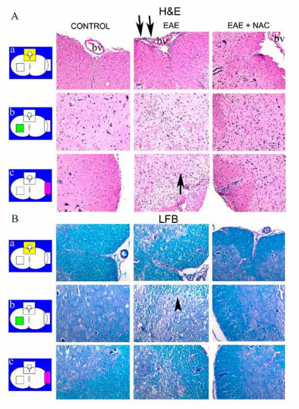
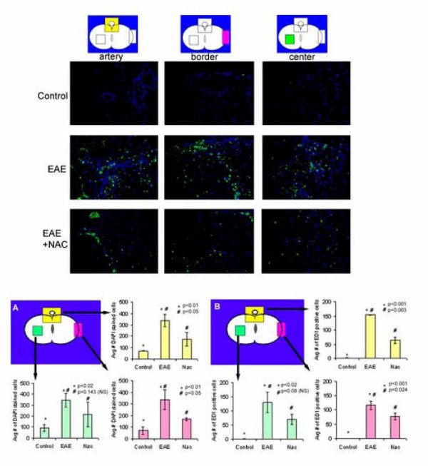
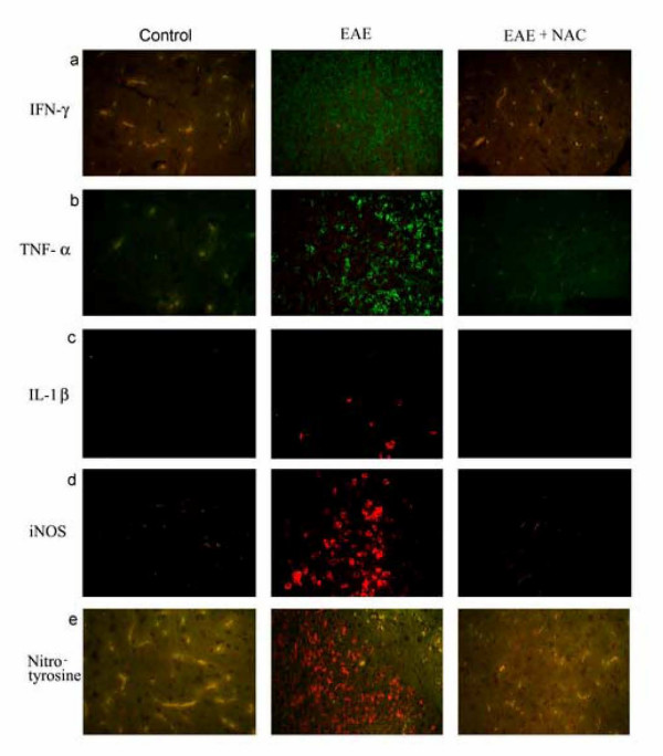
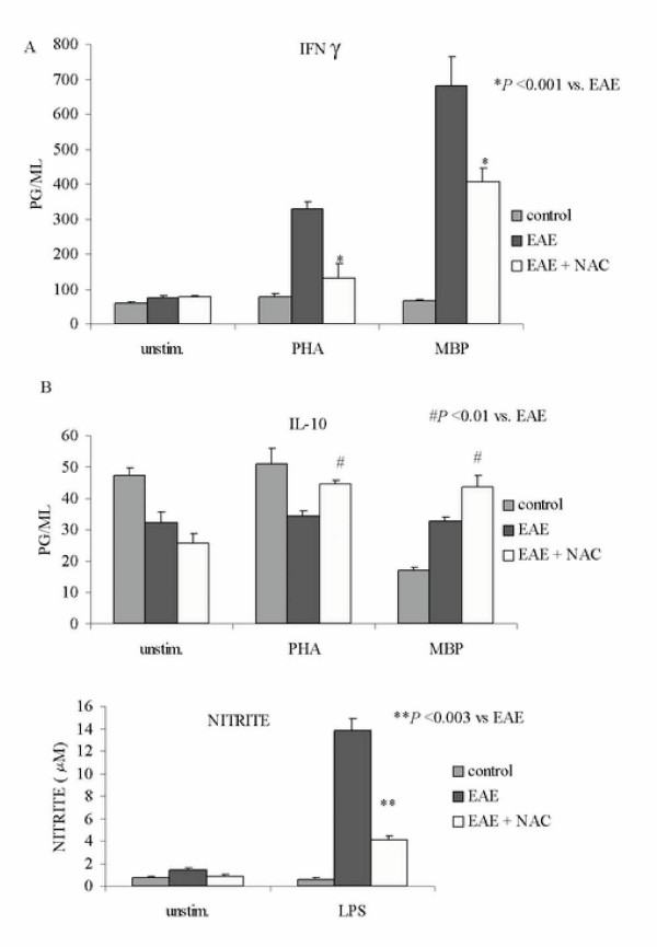
We report that N-acetyl-L-cysteine (NAC) treatment blocked induction of TNF-alpha, IL-1beta, IFN-gamma and iNOS in the CNS and attenuated clinical disease in the myelin basic protein induced model of experimental allergic encephalomyelitis (EAE) in Lewis rats. Infiltration of mononuclear cells into the CNS and induction of inflammatory cytokines and iNOS in multiple sclerosis (MS) and EAE have been implicated in subsequent disease progression and pathogenesis. To understand the mechanism of efficacy of NAC against EAE, we examined its effect on the production of cytokines and the infiltration of inflammatory cells into the CNS. NAC treatment attenuated the transmigration of mononuclear cells thereby lessening the neuroinflammatory disease. Splenocytes from NAC-treated EAE animals showed reduced IFN-gamma production, a Th1 cytokine and increased IL-10 production, an anti-inflammatory cytokine. Further, splenocytes from NAC-treated EAE animals also showed decreased nitrite production when stimulated in vitro by LPS. These observations indicate that NAC treatment may be of therapeutic value in MS against the inflammatory disease process associated with the infiltration of activated mononuclear cells into the CNS.
Figures
Similar articles
-
Lovastatin treatment decreases mononuclear cell infiltration into the CNS of Lewis rats with experimental allergic encephalomyelitis.J Neurosci Res. 2001 Oct 15;66(2):155-62. doi: 10.1002/jnr.1207. J Neurosci Res. 2001. PMID: 11592110
-
Differential tumor necrosis factor alpha expression by astrocytes from experimental allergic encephalomyelitis-susceptible and -resistant rat strains.J Exp Med. 1991 Apr 1;173(4):801-11. doi: 10.1084/jem.173.4.801. J Exp Med. 1991. PMID: 1901078 Free PMC article.
-
Carvacrol ameliorates experimental autoimmune encephalomyelitis through modulating pro- and anti-inflammatory cytokines.Life Sci. 2019 Feb 15;219:257-263. doi: 10.1016/j.lfs.2018.11.051. Epub 2018 Nov 23. Life Sci. 2019. PMID: 30472298
-
Evidence that Fas and FasL contribute to the pathogenesis of experimental autoimmune encephalomyelitis.Arch Immunol Ther Exp (Warsz). 2000;48(5):381-8. Arch Immunol Ther Exp (Warsz). 2000. PMID: 11140465 Review.
-
Cytokines in neuroinflammatory disease: role of myelin autoreactive T cell production of interferon-gamma.J Neuroimmunol. 1992 Oct;40(2-3):211-8. doi: 10.1016/0165-5728(92)90135-8. J Neuroimmunol. 1992. PMID: 1430152 Review.
Cited by
-
Modulation of microglial/macrophage activation by macrophage inhibitory factor (TKP) or tuftsin (TKPR) attenuates the disease course of experimental autoimmune encephalomyelitis.BMC Immunol. 2007 Jul 16;8:10. doi: 10.1186/1471-2172-8-10. BMC Immunol. 2007. PMID: 17634104 Free PMC article.
-
N-Acetylcysteine Suppresses Microglial Inflammation and Induces Mortality Dose-Dependently via Tumor Necrosis Factor-α Signaling.Int J Mol Sci. 2023 Feb 14;24(4):3798. doi: 10.3390/ijms24043798. Int J Mol Sci. 2023. PMID: 36835209 Free PMC article.
-
Analysis of N-acetyl cysteine modified polydimethylsiloxane shunt for improved treatment of hydrocephalus.J Biomed Mater Res B Appl Biomater. 2021 Aug;109(8):1177-1187. doi: 10.1002/jbm.b.34780. Epub 2020 Dec 16. J Biomed Mater Res B Appl Biomater. 2021. PMID: 33331125 Free PMC article.
-
Mitochondrial Oxidative Damage Underlies Regulatory T Cell Defects in Autoimmunity.Cell Metab. 2020 Oct 6;32(4):591-604.e7. doi: 10.1016/j.cmet.2020.07.001. Epub 2020 Jul 31. Cell Metab. 2020. PMID: 32738205 Free PMC article.
-
Intracellular redox state as target for anti-influenza therapy: are antioxidants always effective?Curr Top Med Chem. 2014;14(22):2529-41. doi: 10.2174/1568026614666141203125211. Curr Top Med Chem. 2014. PMID: 25478883 Free PMC article. Review.
References
-
- Markovic-Plese S, McFarland HF. Immunopathogenesis of the multiple sclerosis lesion. Curr Neurol Neurosci Rep. 2001;1:257–262. - PubMed
Grants and funding
LinkOut - more resources
Full Text Sources
