

Bone morphogenetic protein 9 induces the transcriptome of basal forebrain cholinergic neurons
- PMID: 15870197
- PMCID: PMC1088172
- DOI: 10.1073/pnas.0502097102
Bone morphogenetic protein 9 induces the transcriptome of basal forebrain cholinergic neurons
Abstract
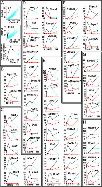
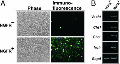
Basal forebrain cholinergic neurons (BFCN) participate in processes of learning, memory, and attention. Little is known about the genes expressed by BFCN and the extracellular signals that control their expression. Previous studies showed that bone morphogenetic protein (BMP) 9 induces and maintains the cholinergic phenotype of embryonic BFCN. We measured gene expression patterns in septal cultures of embryonic day 14 mice and rats grown in the presence or absence of BMP9 by using species-specific microarrays and validated the RNA expression data of selected genes by immunoblot and immunocytochemistry analysis of their protein products. BMP9 enhanced the expression of multiple genes in a time-dependent and, in most cases, reversible manner. The set of BMP9-responsive genes was concordant between mouse and rat and included genes encoding cell-cycle/growth control proteins, transcription factors, signal transduction molecules, extracellular matrix, and adhesion molecules, enzymes, transporters, and chaperonins. BMP9 induced the p75 neurotrophin receptor (NGFR), a marker of BFCN, and Cntf and Serpinf1, two trophic factors for cholinergic neurons, suggesting that BMP9 creates a trophic environment for BFCN. To determine whether the genes induced by BMP9 in culture were constituents of the BFCN transcriptome, we purified BFCN from embryonic day 18 mouse septum by using fluorescence-activated cell sorting of NGFR(+) cells and profiled mRNA expression of these and NGFR(-) cells. Approximately 30% of genes induced by BMP9 in vitro were overexpressed in purified BFCN, indicating that they belong to the BFCN transcriptome in situ and suggesting that BMP signaling contributes to maturation of BFCN in vivo.
Figures

Similar articles
-
Differential modulation of nerve growth factor receptor (p75) and cholinergic gene expression in purified p75-expressing and non-expressing basal forebrain neurons by BMP9.Brain Res. 2008 Dec 30;1246:19-28. doi: 10.1016/j.brainres.2008.09.085. Epub 2008 Oct 14. Brain Res. 2008. PMID: 18952073 Free PMC article.
-
BMP9 (bone morphogenetic protein 9) induces NGF as an autocrine/paracrine cholinergic trophic factor in developing basal forebrain neurons.J Neurosci. 2010 Jun 16;30(24):8221-8. doi: 10.1523/JNEUROSCI.5611-09.2010. J Neurosci. 2010. PMID: 20554873 Free PMC article.
-
BMP9 ameliorates amyloidosis and the cholinergic defect in a mouse model of Alzheimer's disease.Proc Natl Acad Sci U S A. 2013 Nov 26;110(48):19567-72. doi: 10.1073/pnas.1319297110. Epub 2013 Nov 11. Proc Natl Acad Sci U S A. 2013. PMID: 24218590 Free PMC article.
-
The cholinergic system, nerve growth factor and the cytoskeleton.Behav Brain Res. 2011 Aug 10;221(2):515-26. doi: 10.1016/j.bbr.2010.02.024. Epub 2010 Feb 16. Behav Brain Res. 2011. PMID: 20170684 Review.
-
Bone morphogenetic proteins.Growth Factors. 2004 Dec;22(4):233-41. doi: 10.1080/08977190412331279890. Growth Factors. 2004. PMID: 15621726 Review.
Cited by
-
Bone morphogenetic protein 9 serves a protective role in response to ischemic‑reperfusion in the brain by promoting ERK activation.Mol Med Rep. 2018 Feb;17(2):2845-2852. doi: 10.3892/mmr.2017.8253. Epub 2017 Dec 11. Mol Med Rep. 2018. PMID: 29257291 Free PMC article.
-
TGFbeta signaling in Tribolium: vertebrate-like components in a beetle.Dev Genes Evol. 2008 Apr;218(3-4):203-13. doi: 10.1007/s00427-007-0179-7. Epub 2008 Apr 8. Dev Genes Evol. 2008. PMID: 18392881
-
Subtype-specific neurons from patient iPSCs display distinct neuropathological features of Alzheimer's disease.Cell Regen. 2024 Oct 10;13(1):21. doi: 10.1186/s13619-024-00204-y. Cell Regen. 2024. PMID: 39388038 Free PMC article.
-
Transcriptional responses of cultured rat sympathetic neurons during BMP-7-induced dendritic growth.PLoS One. 2011;6(7):e21754. doi: 10.1371/journal.pone.0021754. Epub 2011 Jul 13. PLoS One. 2011. PMID: 21765909 Free PMC article.
-
Crosstalk among electrical activity, trophic factors and morphogenetic proteins in the regulation of neurotransmitter phenotype specification.J Chem Neuroanat. 2016 Apr;73:3-8. doi: 10.1016/j.jchemneu.2015.12.001. Epub 2015 Dec 12. J Chem Neuroanat. 2016. PMID: 26686293 Free PMC article. Review.
References
-
- Fann, M.-J. & Patterson, P. H. (1994) J. Neurochem. 63, 2074–2079. - PubMed
-
- Lopez-Coviella, I., Berse, B., Krauss, R., Thies, R. S. & Blusztajn, J. K. (2000) Science 289, 313–316. - PubMed
-
- Nonner, D., Barrett, E. F., Kaplan, P. & Barrett, J. N. (2001) J. Neurochem. 77, 691–699. - PubMed
-
- Nonner, D., Panickar, K., Barrett, E. F. & Barrett, J. N. (2004) J. Neurochem. 91, 77–87. - PubMed
-
- Bartus, R. T. (2000) Exp. Neurol. 163, 495–529. - PubMed
Publication types
MeSH terms
Substances
Grants and funding
LinkOut - more resources
Full Text Sources
Other Literature Sources
Molecular Biology Databases
Research Materials
Miscellaneous
