Comparative Study
. 2005 May 17;102(20):7227-32.
doi: 10.1073/pnas.0501536102. Epub 2005 May 3.

A common haplotype in the complement regulatory gene factor H (HF1/CFH) predisposes individuals to age-related macular degeneration

Affiliations
Comparative Study

A common haplotype in the complement regulatory gene factor H (HF1/CFH) predisposes individuals to age-related macular degeneration

Gregory S Hageman et al. Proc Natl Acad Sci U S A. .

Abstract

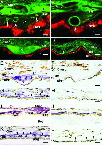
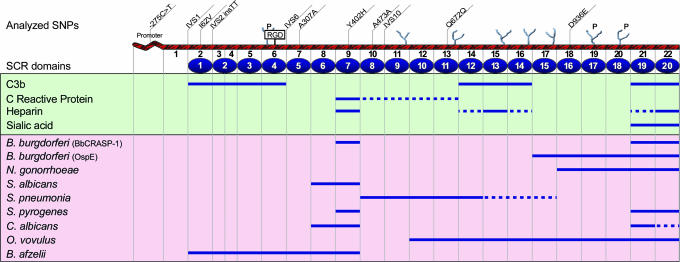
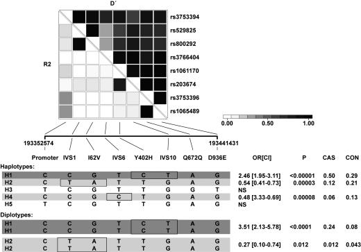
Age-related macular degeneration (AMD) is the most frequent cause of irreversible blindness in the elderly in developed countries. Our previous studies implicated activation of complement in the formation of drusen, the hallmark lesion of AMD. Here, we show that factor H (HF1), the major inhibitor of the alternative complement pathway, accumulates within drusen and is synthesized by the retinal pigmented epithelium. Because previous linkage analyses identified chromosome 1q25-32, which harbors the factor H gene (HF1/CFH), as an AMD susceptibility locus, we analyzed HF1 for genetic variation in two independent cohorts comprised of approximately 900 AMD cases and 400 matched controls. We found association of eight common HF1 SNPs with AMD; two common missense variants exhibit highly significant associations (I62V, chi2 = 26.1 and P = 3.2 x 10(-7) and Y402H, chi2 = 54.4 and P = 1.6 x 10(-13)). Haplotype analysis reveals that multiple HF1 variants confer elevated or reduced risk of AMD. One common at-risk haplotype is present at a frequency of 50% in AMD cases and 29% in controls [odds ratio (OR) = 2.46, 95% confidence interval (1.95-3.11)]. Homozygotes for this haplotype account for 24% of cases and 8% of controls [OR = 3.51, 95% confidence interval (2.13-5.78)]. Several protective haplotypes are also identified (OR = 0.44-0.55), further implicating HF1 function in the pathogenetic mechanisms underlying AMD. We propose that genetic variation in a regulator of the alternative complement pathway, when combined with a triggering event, such as infection, underlie a major proportion of AMD in the human population.


Figures

Fig. 1.
Fig. 1.
Immunolocalization of HF1 (A-H) and MAC (C5b-9) (I-L) in the RPE/choroid (Chor) complex. Ret, retina; Dr, drusen. (A-D) Autofluorescent RPE lipofuscin granules are red (Cy3 channel). (A and B) Confocal immunofluorescence images from an 84-year-old male with atrophic AMD. Anti-HF1 antibody (Advanced Research Technologies, San Diego) labels substructural elements (arrows) in drusen and the subRPE space (green; Cy2 channel). These elements also display IR by using antibodies raised against C3 fragments (iC3b, C3d, and C3dg). The anti-HF1 labeling in the lumens of choroidal capillaries (asterisks) reflects circulating HF1. (Scale bars: A, 5 μm; B, 3 μm.) (C and D) Confocal localization of HF1 in drusen and the subRPE space in an 83-year-old male with AMD (anti-HF1; Quidel, San Diego) (green). (C) Drusen IR is homogeneous. (D) HF1 IR is present throughout the choroid and in the subRPE space (arrowheads). (Scale bars: C, 10 μm; D, 20 μm.) (E-L) Brown pigment in the RPE cytoplasm and choroid is melanin. (E and F) Localization of HF1 in drusen in a 79-year-old donor eye. (E) Anti-HF1 monoclonal antibody-labeling (Quidel) (purple reaction product) is apparent in drusen, along BM, and on the choroidal capillary walls (arrows). (F) Control section of the same eye with no primary antibody. Labeling is absent. (Scale bars: E, 10 μm; F, 10 μm.) (G and H) Localization of HF1 in the macula. (G) Extensive labeling is present along BM, the choroidal capillary walls, and intercapillary pillars (arrows) in a 78-year-old with AMD. (H) Control section from the macula of a donor without AMD; much less labeling is apparent. (Scale bars: G, 20 μm; H, 20 μm.) (I and J) Localization of C5b-9 in the RPE-choroid underlying the macula (I) and extramacula (J) in the same eye of an 81-year-old AMD donor. (I) Intense anti-C5b-9 IR is associated with drusen, BM, and the choroidal capillary endothelium. (J) Outside the macula, there is only sporadic labeling in the vicinity of BM. (Scale bars: I, 20 μm; J, 20 μm.) (K and L) Localization of C5b-9 in the macula from a donor with AMD (K) and from a second donor without AMD (L). (K) Anti-C5b-9 labeling is associated primarily with the choroidal capillary walls (black arrows) and intercapillary pillars (white arrows). Labeling is much more intense in the AMD eye. Note the strong similarity to the anti-HF1 labeling pattern in the macula from the same donor (G). (Scale bars: K, 15 μm; L, 20 μm.)
Fig. 2.
Fig. 2.
Schematic of the HF1/CFH gene. The approximate locations of the 11 SNPs used in the analyses are shown on top of the diagram. The encoded HF1 protein contains 20 SCRs, a RGD domain, and N-linked glycosylation sites (potential sites “P”). Approximate binding sites for pathogens and other substrates are depicted below the diagram based on data published in refs. and . The map is not drawn to scale.
Fig. 3.
Fig. 3.
Association analysis of HF1 haplotypes. A set of the eight most informative SNPs in the HF1 gene were selected and analyzed for pairwise linkage disequilibrium in the Columbia cases and controls. R2 and D′ values are shown. All of the haplotypes with a frequency of >3% are displayed. The ORs for the comparison of cases and controls were calculated, and 95% CIs are shown in brackets. The estimated frequencies of the haplotype in cases (CAS) and controls (CON) are also shown. The major risk haplotype (H1) is shown in dark shading, and the protective haplotypes are shown in light shading. SNPs exclusively found in these risk and protective haplotypes are boxed. Analysis of diplotypes was performed to assess dominant and recessive effects. Homozygotes for the H1 haplotype (H1/H1) are significantly at risk, and H2/H2 individuals are significantly protected. Detailed information pertaining to analyses and access to additional data is provided in Supporting Text: Statistical Analyses.

Comment in

References

    1. Klein, R., Peto, T., Bird, A. & Vannewkirk, M. R. (2004) Am. J. Ophthalmol. 137, 486-495. - PubMed
    1. van Leeuwen, R., Klaver, C. C., Vingerling, J. R., Hofman, A. & de Jong, P. T. (2003) Eur. J. Epidemiol. 18, 845-854. - PubMed
    1. Klein, M. L. & Francis, P. J. (2003) Ophthalmol. Clin. N. Am. 16, 567-574. - PubMed
    1. Tuo, J., Bojanowski, C. M. & Chan, C. C. (2004) Prog. Retinal Eye Res. 23, 229-249. - PMC - PubMed
    1. Klaver, C. C., Wolfs, R. C., Assink, J. J., van Duijn, C. M., Hofman, A. & de Jong, P. T. (1998) Arch. Ophthalmol. 116, 1646-1651. - PubMed

Publication types