Comparative Study
. 2005 May 17;102(20):7145-50.
doi: 10.1073/pnas.0409035102. Epub 2005 May 3.

Visualizing reaction pathways in photoactive yellow protein from nanoseconds to seconds

Affiliations
Comparative Study

Visualizing reaction pathways in photoactive yellow protein from nanoseconds to seconds

Hyotcherl Ihee et al. Proc Natl Acad Sci U S A. .

Abstract

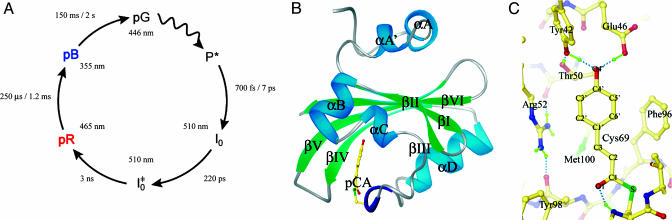
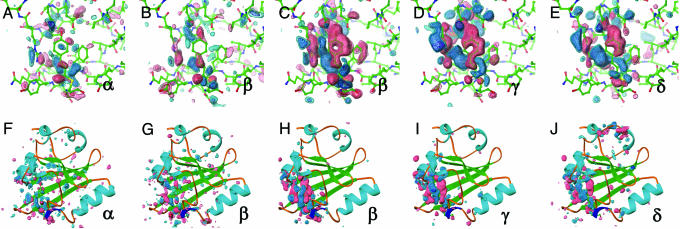
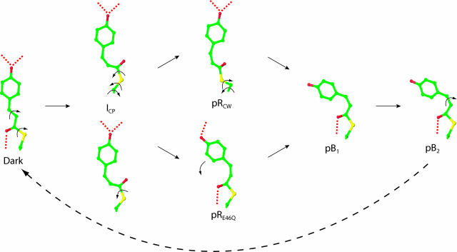
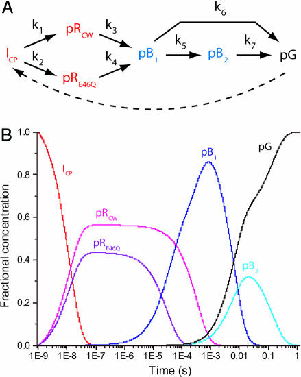
Determining 3D intermediate structures during the biological action of proteins in real time under ambient conditions is essential for understanding how proteins function. Here we use time-resolved Laue crystallography to extract short-lived intermediate structures and thereby unveil signal transduction in the blue light photoreceptor photoactive yellow protein (PYP) from Halorhodospira halophila. By analyzing a comprehensive set of Laue data during the PYP photocycle (forty-seven time points from one nanosecond to one second), we track all atoms in PYP during its photocycle and directly observe how absorption of a blue light photon by its p-coumaric acid chromophore triggers a reversible photocycle. We identify a complex chemical mechanism characterized by five distinct structural intermediates. Structural changes at the chromophore in the early, red-shifted intermediates are transduced to the exterior of the protein in the late, blue-shifted intermediates through an initial "volume-conserving" isomerization of the chromophore and the progressive disruption of hydrogen bonds between the chromophore and its surrounding binding pocket. These results yield a comprehensive view of the PYP photocycle when seen in the light of previous biophysical studies on the system.


Figures

Fig. 1.
Fig. 1.
Overview of PYP (46). (A) The room-temperature PYP photocycle (6, 9). (B) Structure of PYP. The pCA chromophore is shown in yellow; secondary structure is labeled using the notation of Rubinstenn et al. (47). (C) Structure of the chromophore binding pocket of PYP. Hydrogen bonds are shown as green dotted lines and chromophore atoms are labeled according to Borgstahl et al. (5).
Fig. 2.
Fig. 2.
Difference electron density maps for distinct chemical states (α, β, γ, and δ) from an initial kinetic analysis of 47 time points from 1 ns to 1 s. Chromophore-binding pocket (A-E) and whole protein views (F-J) of the difference maps associated with the “α” (A and F) and “β” (B and G) states derived from the ESRF data and from the “β” (C and H), “γ” (D and I), and “δ” (E and J) states from the APS data. Difference maps are contoured at -4σ (red), -3σ (pink), +3σ (cyan), and +4σ (blue). These chemical states are in the order of occurrence in time through the photocycle of PYP. The population of the α, β, γ, and δ states are peaked around at nanoseconds, microseconds, milliseconds, and subseconds time range, respectively (see text and Figs. 3 and 4B).
Fig. 3.
Fig. 3.
Chromophore-binding pocket views of refined intermediate structures and mechanism for the isomerization and rotation of the pCA chromophore upon absorption of blue light. Five distinct structural intermediates (ICP, pRCW, pRE46Q, pB1, and pB2) were identified from four chemical states (α, β, γ, and δ) shown in Fig. 2. ICP is shown twice to demonstrate the biphasic pathways to pRCW and pRE46Q. Isomerization and rotation about single bonds are shown by arrows; hydrogen bonds are dotted. A bicycle pedal mechanism (44), which couples trans-cis isomerization of the C2─C3 double bond with rotation about a nonadjacent single bond, is used for the dark state to ICP transition. Further rotations about single bonds result in the pB1 conformation. pB2 reverts thermally to the dark state with no further detectable intermediates.
Fig. 4.
Fig. 4.
Properties of the chemical kinetic mechanism of the wild-type PYP photocycle. (A) General chemical kinetic mechanism used to fit the data. The dashed arrow indicates the light-driven reaction from pG to the first intermediate observed here, ICP. (B) Predicted concentrations of intermediates [ICP (red), pRCW (magenta), pRE46Q (purple), pB1 (blue), pB2 (cyan), and the dark state (black)] after posterior analysis with rate coefficients (s-1): k1 = 4.8 × 10⁷; k2 = 3.7 × 10⁷; k3 = 3.0 × 10³; k4 = 3.3 × 10⁴; k5 = 55; k6 = 100; and k7 = 7.1. The seven rate coefficients correspond to time constants of 21 ns, 27 ns, 333 μs, 30 μs, 18 ms, 10 ms, and 141 ms, respectively.

References

    1. Sprenger, W. W., Hoff, W. D., Armitage, J. P. & Hellingwerf, K. J. (1993) J. Bacteriol. 175, 3096-3104. - PMC - PubMed
    1. Hellingwerf, K. J., Hendriks, J. & Gensch, T. (2003) J. Phys. Chem. A 107, 1082-1094.
    1. Cusanovich, M. A. & Meyer, T. E. (2003) Biochemistry 42, 4759-4770. - PubMed
    1. Taylor, B. L. & Zhulin, I. B. (1999) Microbiol. Mol. Biol. Rev. 63, 479-506. - PMC - PubMed
    1. Borgstahl, G. E., Williams, D. R. & Getzoff, E. D. (1995) Biochemistry 34, 6278-6287. - PubMed

Publication types