. 2005 May 15;19(10):1162-74.
doi: 10.1101/gad.1291305. Epub 2005 May 3.

PPM1D dephosphorylates Chk1 and p53 and abrogates cell cycle checkpoints

Affiliations

PPM1D dephosphorylates Chk1 and p53 and abrogates cell cycle checkpoints

Xiongbin Lu et al. Genes Dev. .

Abstract

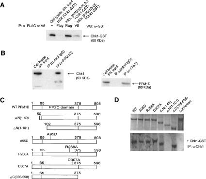
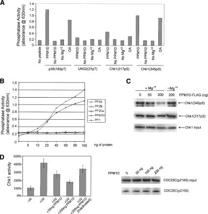
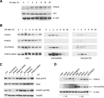
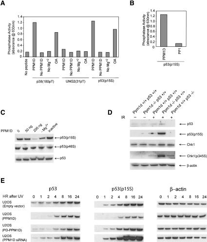
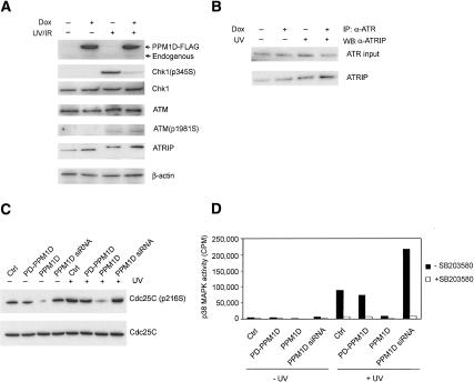
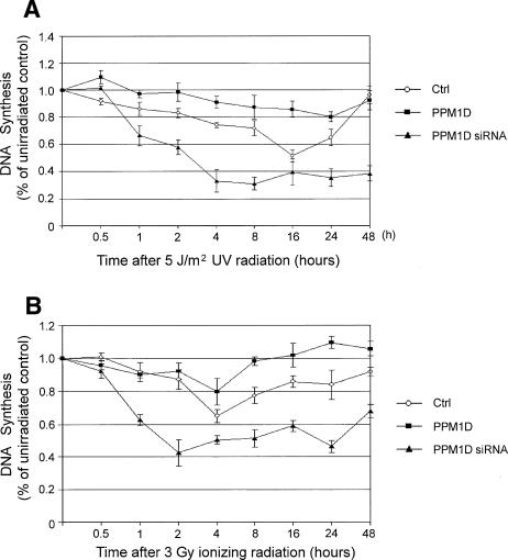
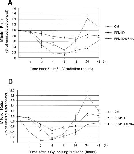
The ATM (ataxia-telangiectasia mutated) and ATR (ataxia-telangiectasia and Rad3-related) kinases respond to DNA damage by phosphorylating cellular target proteins that activate DNA repair pathways and cell cycle checkpoints in order to maintain genomic integrity. Here we show that the oncogenic p53-induced serine/threonine phosphatase, PPM1D (or Wip1), dephosphorylates two ATM/ATR targets, Chk1 and p53. PPM1D binds Chk1 and dephosphorylates the ATR-targeted phospho-Ser 345, leading to decreased Chk1 kinase activity. PPM1D also dephosphorylates p53 at phospho-Ser 15. PPM1D dephosphorylations are correlated with reduced cellular intra-S and G2/M checkpoint activity in response to DNA damage induced by ultraviolet and ionizing radiation. Thus, a primary function of PPM1D may be to reverse the p53 and Chk1-induced DNA damage and cell cycle checkpoint responses and return the cell to a homeostatic state following completion of DNA repair. These homeostatic functions may be partially responsible for the oncogenic effects of PPM1D when it is amplified and overexpressed in human tumors.


Figures

Figure 1.
Figure 1.
PPM1D binds Chk1. (A) Immunoprecipitation Western analysis shows PPM1D–Chk1 interaction. Chk1-GST and PPM1D tagged with V5 or Flag were introduced into HEK cells and lysates immunoprecipitated with anti-Flag or V5 prior to Western blot probing with a GST antibody. (B) Endogenous PPM1D interacts with endogenous Chk1. (Left panel) Immunoprecipitation of lysates from U2OS cells with anti-PPM1D or control antibody followed by Western blot probing with anti-Chk1 antibody shows that endogenous Chk1 is detected on blots containing immunoprecipitated PPM1D, but not following immunoprecipitation with control antibody. (Right panel) A reciprocal experiment utilizing immunoprecipitation with an anti-Chk1 antibody followed by Western blot probing of the immunoprecipitated lysate with an anti-PPM1D antibody confirmed the endogenous Chk1–PPM1D interaction. (C) Diagram of mutant forms of PPM1D constructed to determine Chk1-binding domains. Below the wild-type protein at the top is shown two N-terminal deletion mutants, three phosphatase-dead point mutants, and one C-terminal truncation mutant. The conserved phosphatase domain of PPM1D extends from amino acid 65 to 375. (D) Chk1 binds to mutant forms of PPM1D except for one mutant missing part of the phosphatase domain. Recombinant GST-Chk1 was incubated with in vitro translated and 35S-labeled wild-type or mutant PPM1D (upper panel) followed by immunoprecipitation with Chk1 antibodies prior to SDS–polyacrylamide gel electrophoresis and autoradiography (lower panel). Only the PPM1D mutant ΔN(1–101) missing key elements of the conserved phosphatase domain did not bind Chk1.
Figure 2.
Figure 2.
PPM1D dephosphorylates Chk1 phospho-Ser 345 and inhibits Chk1 kinase activity in vitro. (A) PPM1D dephosphorylates Chk1 phospho-Ser 345. Phosphopeptides from p38 MAP kinase (positive control), UNG2 (negative control), and Chk1 (phospho-Ser 317 or phospho-Ser 345) were incubated with PPM1D in an in vitro phosphatase assay. Release of free phosphate was determined by absorbance at 630 nm in the presence of molybdate dye. Reactions were also performed in the absence of magnesium or peptide or in the presence of okadaic acid. (B) PPM1D and PP1, but not other serine/threonine phosphatases, exhibit in vitro phosphatase activity on a Chk1 phosphopeptide. Indicated amounts of purified serine/threonine phosphatases PPM1D, PP1, PP2A, PP2B, and PP2C α were incubated with Chk1 Ser 345 phosphopeptide in an in vitro phosphatase assay under conditions similar to those in panel A. (C) PPM1D dephosphorylates intact Chk1 at phosphoSer 345. An in vitro phosphatase assay was performed by incubating immunoprecipitated Chk1 and purified PPM1D prior to Western blot probing with antibodies to Chk1 phospho-Ser 345 and phospho-Ser 317. (D) PPM1D inhibits Chk1 kinase activity in vitro. Chk1 immunopurified from untreated or UV-treated U2OS cells was incubated with active or inactive PPM1D prior to incubation with a Chk1 target peptide (Ser 216 from CDC25C). (Left panel) Chk1 kinase activity was measured by 32P labeling of substrate peptide. (Right panel) The dephosphorylation of the CDC25C target peptide was not a result of direct PPM1D dephosphorylation because when increasing amounts of purified PPM1D were incubated only with immunopurified CDC25C in the absence of Chk1, no decrease in CDC25C Ser 216 phosphorylation was noted when the Western blot was probed with a CDC25C Ser 216 phosphospecific antibody.
Figure 3.
Figure 3.
PPM1D inhibits phosphorylation of Chk1 and Chk1 targets in cells. (A) Time course of PPM1D protein expression following UV irradiation. U2OS cells were transfected with empty vector and were UV radiated at 30 J/m2. Cell lysates were harvested at varying time points (0–24 h) after UV treatment and subjected to Western blot probing with anti-PPM1D and anti-PP1 antibody. (B) PPM1D inhibits Chk1 phosphorylation in cells following UV irradiation. U2OS cells were untransfected or transfected with wild-type PPM1D or phosphatase-dead (PD) constructs or with PPM1D siRNA, then UV irradiated and harvested before or at varying time points after UV treatment. Lysates were Western blot probed with antibodies to Chk1 protein and Chk1 phospho-Ser 345 and phospho-Ser 317. (C) Phosphorylation of downstream Chk1 targets is inhibited by PPM1D. Lysates from U2OS cells were transfected, UV irradiated or unirradiated as in B, and analyzed by Western blot for Cdc2 Tyr 15, Cdc2 protein, CDC25C phospho-Ser 216, and CDC25C protein. (D) Breast cancer cells with amplified PPM1D exhibit attenuated UV-induced Chk1 phosphorylation. Control cell lines (HEK and U2OS) and breast cancer cell lines with amplified PPM1D (MCF-7, BT474, and MDAMB231) were unirradiated or UV irradiated (30 J/m2), and lysates were harvested 2 h later, prior to blot probing with antibodies to PPM1D and Chk1 phospho-Ser 345.
Figure 4.
Figure 4.
p53 Ser 15 is dephosphorylated by PPM1D. (A) PPM1D dephosphorylates p53 Ser 15 phosphopeptide in vitro. p53 Ser 15 phosphopeptide was incubated with purified PPM1D in an in vitro phosphatase assay. Reactions were also performed in the absence of magnesium or peptide or in the presence of okadaic acid. Positive (p38 180pT) and negative (UNG2 31pT) control phosphopeptides were also assessed for PPM1D activity. (B) PPM1D, but not PP1, has in vitro phosphatase activity on the p53 Ser 15 phosphopeptide. Assay conditions were similar to those in panel A. (C) PPM1D dephosphorylates intact p53 at Ser 15. Immunopurified p53 from UV-irradiated U2OS cells was incubated with PPM1D in an in vitro phosphatase assay and was Western blot probed with antibodies to p53 phospho-Ser 15, p53 phospho-Ser 46, and p53 protein. (D) PPM1D null fibroblasts exhibit increased p53 Ser 15 phosphorylation after IR. Lysates harvested from mouse embryo fibroblasts of the designated PPM1D and p53 genotypes 2 h after treatment with 5 Gy IR were Western blot probed with antibodies to p53 protein (first panel), p53 phospho-Ser 15 (second panel), Chk1 protein (third panel), Chk1 phospho-Ser 345 (fourth panel), and β-actin (fifth panel). (E) PPM1D inhibits p53 phosphorylation at Ser 15 in UV-treated cells. U2OS cells were untransfected or transfected with wild-type PPM1D or phosphatase-dead (PD) PPM1D or PPM1D siRNA, then UV or mock irradiated and harvested before or at varying times after 30 J/m2 UV treatment. Cell lysates were Western blot probed with antibodies to p53 protein (left panel), p53 phospho-Ser 15 (center panel), or β-actin (right panel).
Figure 5.
Figure 5.
Effects of PPM1D on cellular stress and checkpoint targets. (A) ATM and ATRIP damage-induced post-translational modifications appear to be unaffected by PPM1D. U2OS tet on (PPM1D) cells were incubated with or without doxycycline prior to treatment with 5 Gy IR (Chk1, ATM, β-actin panels) or 30 J/m2 UV radiation (ATRIP panel). Two hours after irradiation, lysates were harvested and subjected to SDS–polyacrylamide gel electrophoresis and Western blot probing with antibodies to PPM1D (top panel), Chk1 phospho-Ser 345 (second panel), Chk1 protein (third panel), ATM protein (fourth panel), ATM phospho-Ser 1981 (fifth panel), ATRIP protein (sixth panel), and β-actin (seventh panel). (B) ATR–ATRIP interactions appear to be unaffected by PPM1D. U2OS tet on (PPM1D) cells were incubated with or without doxycycline and mock treated or treated with 30 J/m2 UV radiation. Lysates were harvested 2 h after irradiation and subjected to immunoprecipitation with an anti-ATR antibody followed by Western blotting with antibodies to ATR (top panel) or ATRIP (bottom panel). (C) PPM1D overexpression leads to CDC25C dephosphorylation in the absence of p38 MAP kinase signaling. U2OS cells were incubated in the presence of the p38 MAP kinase inhibitor SB203580 and transfected with empty vector, phosphatase-dead PPM1D, wild-type PPM1D, or PPM1D siRNA prior to UV or mock treatment. Lysates were immunoprecipitated with antibody specific to the Ser 216 phosphorylated form of CDC25C. (D) MAP kinase inhibitor SB203580 completely abrogates p38 MAP kinase activity. U2OS cells were transfected and UV or mock treated as in panel A and lysates were measured for p38 activity by assessment of p38 target MAPKAP-K2 kinase activity on a target peptide.
Figure 6.
Figure 6.
PPM1D abrogates the intra-S-phase checkpoint in response to UV and IR. (A) PPM1D inhibits the intra-S checkpoint in response to UV radiation. U2OS tet on (PPM1D) cells were irradiated with 5 J/m2 UV radiation, incubated for varying lengths of time (0.5–48 h), pulse labeled with 3H-thymidine for 15 min, and harvested, and cell lysates were measured for 3H-thymidine incorporation compared with unirradiated control cells. The cells were either maintained under normal conditions in the absence of doxycycline after transfection with control siRNA (Ctrl, circles), under PPM1D overexpression conditions in the presence of doxycycline and control siRNA (PPM1D, squares), or under reduced PPM1D expression (without doxycycline) after transfection of PPM1D siRNA (PPM1D siRNA, triangles). Values are expressed as relative incorporation of 3H-thymidine compared with mock irradiated control cells. (B) PPM1D inhibits the intra-S-phase checkpoint in response to IR. The experiments were carried out exactly as in panel A, except that all cells were treated with 3 Gy IR instead of UV radiation.
Figure 7.
Figure 7.
PPM1D abrogates the G2/M checkpoint in response to UV and IR. (A) PPM1D inhibits the G2/M checkpoint in response to UV radiation. U2OS tet on (PPM1D) cells were irradiated with 5 J/m2 UV radiation, incubated for varying lengths of time (0.5–48 h), harvested, fixed, and incubated with antiphosphohistone H3 antibody, followed by FITC-conjugated secondary antibody and fluorescence measurement by flow cytometry. The cells were maintained under normal conditions in the absence of doxycycline after transfection with control siRNA (Ctrl, circles), under PPM1D overexpression conditions in the presence of doxycycline and control siRNA (PPM1D, squares), or under reduced PPM1D expression (without doxycycline) after transfection of PPM1D siRNA (PPM1D siRNA, triangles). Values are expressed as relative phosphohistone H3 fluorescence compared with mock irradiated control cells. (B) PPM1D inhibits the G2/M-phase checkpoint in response to IR. The experiments were carried out exactly as in panel A, except that all cells were treated with 3 Gy IR instead of UV radiation.

References

    1. Ali A., Zhang, J., Bao, S., Liu, I., Otterness, D., Dean, N.M., Abraham, R.T., and Wang, X.F. 2004. Requirement of protein phosphatase 5 in DNA-damage-induced ATM activation. Genes & Dev. 18: 249–254. - PMC - PubMed
    1. Bakkenist C.J. and Kastan, M.B. 2003. DNA damage activates ATM through intermolecular autophosphorylation and dimer dissociation. Nature 421: 499–506. - PubMed
    1. ____. 2004a. Initiating cellular stress responses. Cell 118: 9–17. - PubMed
    1. ____. 2004b. Phosphatases join kinases in DNA-damage response pathways. Trends Cell Biol. 14: 339–341. - PubMed
    1. Banin S., Moyal, L., Shieh, S., Taya, Y., Anderson, C.W., Chessa, L., Smorodinsky, N.I., Prives, C., Reiss, Y., Shiloh, Y., et al. 1998. Enhanced phosphorylation of p53 by ATM in response to DNA damage. Science 281: 1674–1677. - PubMed

Publication types

MeSH terms