. 2005 May 15;19(10):1238-48.
doi: 10.1101/gad.1309605. Epub 2005 May 3.

RNA-guided RNA modification: functional organization of the archaeal H/ACA RNP

Affiliations

RNA-guided RNA modification: functional organization of the archaeal H/ACA RNP

Daniel L Baker et al. Genes Dev. .

Abstract

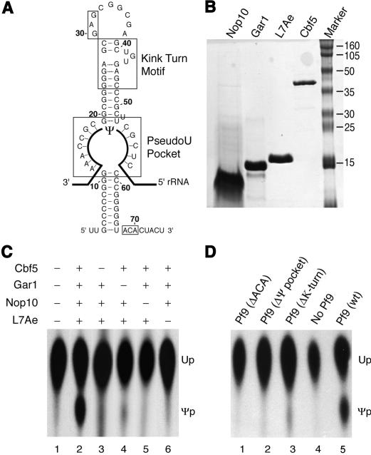
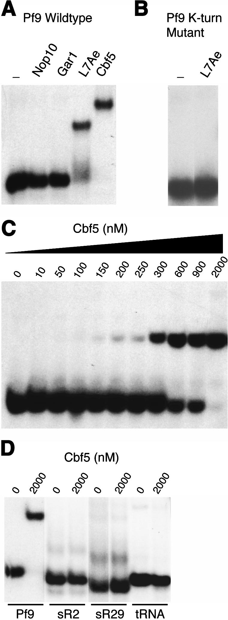
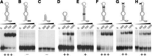
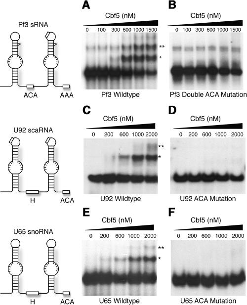
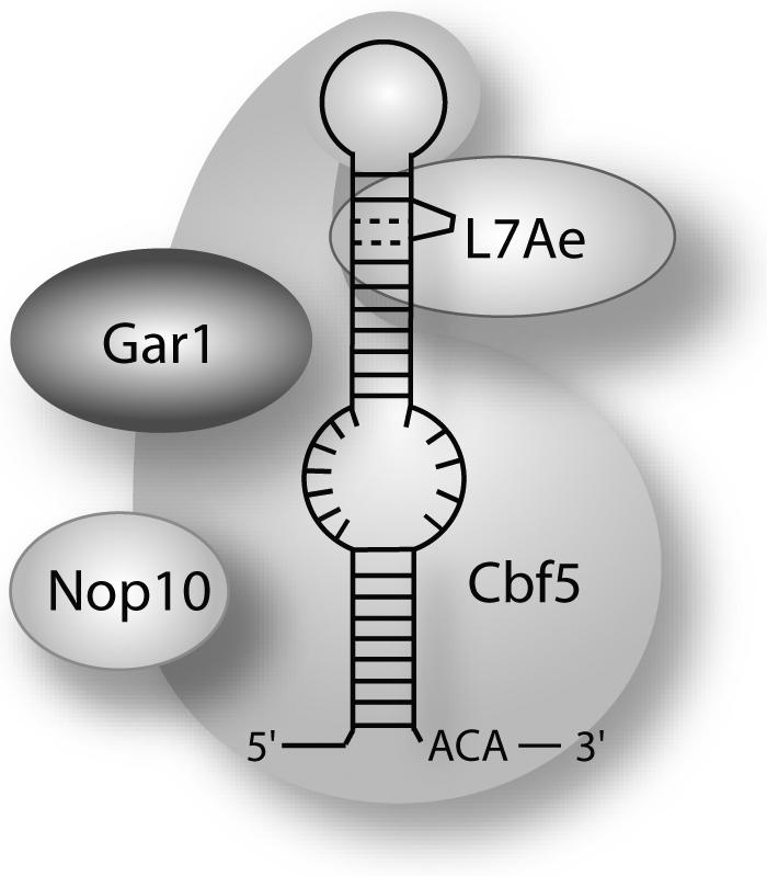
In eukaryotes and archaea, uridines in various RNAs are converted to pseudouridines by RNA-guided RNA modification complexes termed H/ACA RNPs. Guide RNAs within the complexes base-pair with target RNAs to direct modification of specific ribonucleotides. Cbf5, a protein component of the complex, likely catalyzes the modification. However, little is known about the organization of H/ACA RNPs and the roles of the multiple proteins thought to comprise the complexes. We have reconstituted functional archaeal H/ACA RNPs from recombinant components, defined the components necessary and sufficient for function, and determined the direct RNA-protein and protein-protein interactions that occur between the components. The results provide substantial insight into the functional organization of this RNP. The functional complex requires a guide RNA and each of four proteins: Cbf5, Gar1, L7Ae, and Nop10. Two proteins interact directly with the guide RNA: L7Ae and Cbf5. L7Ae does not interact with other H/ACA RNP proteins in the absence of the RNA. We have defined two novel functions for Cbf5. Cbf5 is the protein that specifically recognizes and binds H/ACA guide RNAs. In addition, Cbf5 recruits the two other essential proteins, Gar1 and Nop10, to the pseudouridylation guide complex.


Figures

Figure 1.
Figure 1.
Reconstitution of functional pseudouridylation guide RNPs from recombinant RNA and protein components. (A) Sequence and secondary structure of Pf9 H/ACA guide RNA with important elements indicated. Box ACA is located at the base of the hairpin structure near the 3′ end of the RNA (nucleotides 68–70). The pseudouridylation pocket is an internal loop bounded by the upper and lower stems of the hairpin. The nucleotides within the pocket base-pair with the rRNA substrate (represented as solid bold line), positioning the unpaired uridine to be modified (ψ) at the top of the loop. A kink-turn motif is located in the upper stem, near the terminal loop of the hairpin and consists of an asymmetric loop containing two G-A base pairs and flanked by two short stems (Klein et al. 2001). A GAG sequence present in the terminal loop of Pf9 and other archaeal H/ACA RNAs is indicated (nucleotides 30–32). (B) Purified samples of H/ACA RNP proteins Cbf5, Gar1, Nop10, and L7Ae analyzed by SDS PAGE and Coomassie protein staining are shown. (C) Pseudouridylation activity of various combinations of the four recombinant H/ACA RNP proteins. Pf9 guide RNA and substrate RNA (containing a single, 32P-labeled target uridine) were incubated with the indicated combinations of proteins. Pseudouridylation was assessed by TLC separation of nucleotides (obtained by nuclease P1 digestion of RNA) under established conditions where pseudouridine (ψp) migrates more slowly than uridine (Up) (Yu et al. 2001). Autoradiographs of TLC plates are shown. (D) Effect of mutations in Pf9 guide RNA on pseudouridylation activity. Box ACA was mutated to UGU (ΔACA). Pseudouridylation pocket was eliminated by replacement of sequence on one side of the loop with sequence complementary to other side of the loop (Δψ pocket). The K-turn was mutated by disruption of critical GA base pairs (substitution of GA with CC; ΔK-turn). The indicated mutant or wild-type Pf9 guide RNA was incubated with the four recombinant proteins and substrate RNA, and pseudouridylation activity was assessed as in C.
Figure 2.
Figure 2.
Cbf5 interacts directly and specifically with Pf9 H/ACA guide RNA. Direct interactions of proteins with 32P-labeled RNAs were investigated by native gel mobility shift analysis and autoradiography. (A) Pf9 RNA was incubated with each of the four recombinant H/ACA RNP proteins or no protein (–). (B) The K-turn of Pf9 was disrupted and the mutant RNA was incubated with L7Ae. (C) Wild-type Pf9 was incubated with increasing concentrations of Cbf5 (0–2000 nM) to assess the apparent Kd of the observed interaction. (D) Cbf5 was incubated with non-H/ACA RNAs including P. furiosus C/D RNAs sR2 and sR29 and a human tRNA to assess the specificity of the observed interaction.
Figure 3.
Figure 3.
Elements of the H/ACA guide RNA important for Cbf5 interaction. The ability of Cbf5 to interact with mutants and fragments of Pf9 was assessed by native gel mobility shift analysis with a range of concentrations of Cbf5. Each panel shows a diagram of the RNA tested (location of mutations indicated with X), autoradiograph of gel shift analysis, and scaled estimate of the extent of interaction relative to wild-type Pf9 (– to +++). (A) Wild-type Pf9. (B) Mutation of box ACA (to UGU). (C) Deletion of the terminal loop, K-turn, upper stem, and pseudouridylation pocket. (D) Deletion of the terminal loop, K-turn, and upper stem. (E) Closure of pseudouridylation pocket by replacement of sequence on one side of the loop with sequence complementary to other side of the loop. (F) Disruption of critical GA base pairs in K-turn by substitution of GA with CC. (G) Replacement of terminal loop with tetra-loop. (H) Mutation of GAG in terminal loop (to CUC).
Figure 4.
Figure 4.
Cbf5 also interacts with archaeal and eukaryotic double hairpin H/ACA RNAs. (A,C,E) The ability of Cbf5 to interact with double hairpin H/ACA RNAs Pf3 (a P. furiosus guide RNA), U92 (a eukaryotic scaRNA), and U65 (a eukaryotic snoRNA) was assessed by native gel mobility shift analysis with a range of concentrations of Cbf5. Distinct RNP complexes are indicated with single and double asterisks. A diagram of the RNA tested is shown to the left of each panel. (B,D,F) In order to assess the specificity of the observed interactions and importance of box ACA, native gel mobility shift analysis was performed with RNAs in which the box ACA elements were mutated (ACA to UGU, or AAA to UUU in the case of the 3′ element of Pf3).
Figure 5.
Figure 5.
Cbf5 interacts with Gar1 and Nop10 to form a heterotrimeric protein complex. Combinations of the four H/ACA RNP proteins (indicated as C [Cbf5], L [L7Ae], G [Gar1], and N [Nop10]) were incubated in approximately equimolar amounts (I [input] lanes). In each panel the his-tagged protein is designated with an asterisk. Bovine serum albumin (BSA) was also added to the protein mixtures. The his-tagged proteins were purified using nickel agarose resin. Input (I lanes) and bound (B lanes) samples were compared following 15% Tris-tricine gel electrophoresis and Coomassie blue staining.
Figure 6.
Figure 6.
Assembly of H/ACA RNP proteins with an H/ACA guide RNA. 32P-labeled wild-type (wt) or ACA mutant (ΔACA) Pf9 RNAs were incubated with one or more of the four proteins as indicated. The resultant RNP complexes were detected by native gel shift analysis followed by autoradiography. The distinct complex formed in the presence of all four proteins is indicated with an asterisk.
Figure 7.
Figure 7.
Organization of an archaeal pseudouridylation guide RNP complex. The results of this study suggest the model that is shown. L7Ae interacts directly with the K-turn of the guide RNA. Cbf5 also interacts directly and independently with the guide RNA, making extensive contacts that may include box ACA, the pseudouridylation pocket, and the terminal loop. Association of Gar1 and Nop10 with the complex is mediated by their individual interactions with Cbf5. Close contacts between the various components may occur in the context of the assembled RNP, but no evidence of additional independent interactions was obtained in this study.

Similar articles

Cited by

References

    1. Aittaleb M., Rashid, R., Chen, Q., Palmer, J.R., Daniels, C.J., and Li, H. 2003. Structure and function of archaeal box C/D sRNP core proteins. Nat. Struct. Biol. 10: 256–263. - PubMed
    1. Ambros V. 2004. The functions of animal microRNAs. Nature 431: 350–355. - PubMed
    1. Aravind L. and Koonin, E.V. 2001. THUMP—A predicted RNA-binding domain shared by 4-thiouridine, pseudouridine synthases and RNA methylases. Trends Biochem. Sci. 26: 215–217. - PubMed
    1. Bachellerie J.-P. and Cavaille, J. 1998. Small nucleolar RNAs guide the ribose methylations of eukaryotic rRNAs. In Modification and editing of RNA (eds. H. Grosjean and B. Benne), pp. 255–272. ASM Press, Washington, DC.
    1. Balakin A.G., Smith, L., and Fournier, M.J. 1996. The RNA world of the nucleolus: Two major families of small RNAs defined by different box elements with related functions. Cell 86: 823–834. - PubMed

Publication types

MeSH terms