. 2005 May;25(10):3886-95.
doi: 10.1128/MCB.25.10.3886-3895.2005.

Regulation of the deubiquitinating enzyme CYLD by IkappaB kinase gamma-dependent phosphorylation

Affiliations

Regulation of the deubiquitinating enzyme CYLD by IkappaB kinase gamma-dependent phosphorylation

William Reiley et al. Mol Cell Biol. 2005 May.

Abstract

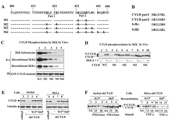
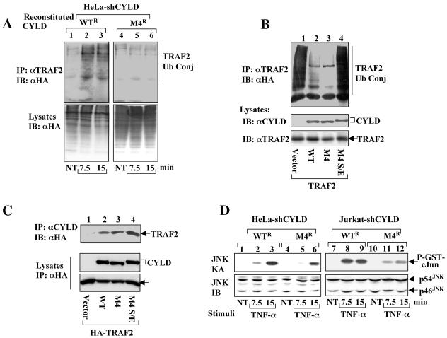
Tumor suppressor CYLD is a deubiquitinating enzyme (DUB) that inhibits the ubiquitination of key signaling molecules, including tumor necrosis factor (TNF) receptor-associated factor 2 (TRAF2). However, how the function of CYLD is regulated remains unknown. Here we provide evidence that inducible phosphorylation of CYLD is an important mechanism of its regulation. Under normal conditions, CYLD dominantly suppresses the ubiquitination of TRAF2. In response to cellular stimuli, CYLD undergoes rapid and transient phosphorylation, which is required for signal-induced TRAF2 ubiquitination and activation of downstream signaling events. Interestingly, the CYLD phosphorylation requires IkappaB kinase gamma (IKKgamma) and can be induced by IKK catalytic subunits. These findings suggest that CYLD serves as a novel target of IKK and that the site-specific phosphorylation of CYLD regulates its signaling function.


Figures

FIG. 1.
FIG. 1.
CYLD knockdown results in constitutive ubiquitination of TRAF2. 293 (A) or HeLa (B) cells were transfected using Lipofectamine 2000 with control or CYLD-specific siRNA together with pcDNA-HA-ubiquitin. The cells were stimulated with TNF-α (50 ng/ml) and then lysed in a buffer containing inhibitors of ubiquitin hydrolases. Endogenous TRAF2 was isolated by IP using anti-TRAF2 antibody, and the ubiquitinated TRAF2 was detected by IB using an HRP-conjugated anti-HA antibody (top panel). The intracellular level of CYLD and TRAF2 was analyzed by IB using anti-CYLD and anti-TRAF2 antibodies (middle and bottom panels). M, molecular mass. NT, not treated.
FIG. 2.
FIG. 2.
CYLD is phosphorylated in response to diverse cellular stimuli. (A) Jurkat T cells were stimulated with either T-cell mitogens (50 ng/ml of PMA plus 1 μM of ionomycin) or TNF-α (20 ng/ml) for the indicated times. To prevent loss of the phosphorylated CYLD, the cells were lysed in a kinase lysis buffer supplemented with phosphatase inhibitors. CYLD proteins were concentrated by IP (using anti-CYLD) and then fractionated in low-percentage (6%) SDS gels in order to separated the basal and phosphorylated CYLD bands. In lanes 11 to 13, the CYLD immune precipitates isolated from TNF-α-stimulated cells (15 min) were either left on ice (NT) or incubated at 37°C for 30 min in calf intestinal alkaline phosphatase (CIP; lane 13) or buffer control (lane 12). The phosphorylated (P-CYLD) and basal (CYLD) forms of CYLD were detected by IB using anti-CYLD (upper panel). The cell lysates were also subjected to IB to detect IκBα degradation (lower panel). (B) Immune complex kinase assays to detect the activation of IKK. Jurkat cells were stimulated with PMA plus inomycin (Iono) as described for panel A. IKK complex was isolated by IP using anti-IKKγ antibody and subjected to kinase assays using GST-IκBα(1-54) as a substrate. The phosphorylated substrate (P-GST-IκBα) is indicated. (C to E) CYLD phosphorylation (upper panel) and IκBα degradation (lower panel) were analyzed in TNF-α-stimulated 293 and HeLa cells and LPS-stimulated BJAB B cells as described for panel A.
FIG. 3.
FIG. 3.
CYLD phosphorylation is mediated by IKK. (A) Inducible phosphorylation of CYLD requires IKKγ. Parental Jurkat cells, IKKγ-deficient Jurkat mutant (JM4.5.2), and IKKγ-reconstituted JM4.5.2 cells were either not treated (−) or were stimulated with PMA plus ionomycin for 15 min. Cell lysates were subjected to CYLD phosphorylation and IκBα degradation analyses as described for Fig. 2A. (B) CYLD phosphorylation by transfected IKK. 293 cells were transfected with HA-tagged CYLD together with either empty vector or expression vectors encoding IKKβ (0.5 μg), IKKα (0.5 μg), or IKKβ (0.5 μg) plus IKKγ (25 ng). CYLD phosphorylation was analyzed by IB using anti-HA antibody. (C) Phosphorylation of CYLD truncation mutants. CYLD truncation mutants covering different lengths of its C terminus (indicated by the amino acid numbers) were expressed in 293 cells in either the absence (−) or presence (+) of HA-IKKβ plus HA-IKKγ. Phosphorylation of CYLD (upper panel) and expression of IKKβ (middle panel) and IKKγ (bottom panel) were analyzed by IB using anti-HA. Phosphorylated CYLD bands are indicated by an arrowhead. (D) CYLD/IKKγ physical interaction. Full-length (FL) or truncated forms of CYLD (tagged with HA) were coexpressed with myc-tagged IKKγ. The IKKγ complex was isolated by IP using anti-myc followed by detecting the associated CYLD proteins by IB using anti-HA-HRP (upper panel). The expression level of IKKγ was analyzed by IB using anti-myc (lower panel). Lane 1 is a negative control that was transfected with CYLD only. (E) In vitro kinase assays to demonstrate CYLD phosphorylation by IKK homoenzyme. IKK holoenzyme was isolated by IP (using anti-IKKγ) from untreated (NT), PMA-ionomycin-stimulated (7.5 min), or TNF-α-stimulated (7.5 min) Jurkat cells and subjected to in vitro kinase assays using GST-IκBα(1-54) (lower panel) or GST-CYLD(403-513) (upper panel) as a substrate. (F) In vitro kinase assays were performed using IKK complex isolated from PMA-ionomycin-stimulated Jurkat cells and GST-CYLD(403-513) or GST substrate. GST-CYLD(403-513), but not GST, was phosphorylated by IKK. (G) CYLD phosphorylation by recombinant IKKβ. In vitro kinase assays were performed using the indicated amounts of purified IKKβ recombinant protein and GST-IκBα(1-54) (lanes 1 to 5) or GST-CYLD(403-513) (lanes 6 to 10) substrate. Autophosphorylated IKKβ (P-IKKβ) and phosphorylated substrates are indicated.
FIG. 4.
FIG. 4.
Site-specific phosphorylation of CYLD by IKK. (A) Amino acid sequence of CYLD phosphorylation region. The putative phosphorylation sites (serines) are boldface, and two serine pairs are indicated. (B) Sequence homology between the CYLD serine pairs and the IKK phosphorylation sites within IκBα and IκBβ. (C) Phosphorylation of CYLD mutants by IKK holoenzyme (top panel) and recombinant IKKs (middle panels). GST-CYLD(403-513) with a wild-type phosphorylation site (WT) or the various mutations (indicated in panel A) were subjected to in vitro kinase assays (KA) using IKK holoenzyme isolated from mitogen-stimulated Jurkat cells (top panel), recombinant IKKβ (second panel), or recombinant IKKα (third panel). The substrate amounts were monitored by IB using anti-GST (bottom panel). (D) In vivo phosphorylation of CYLD mutants by IKK. HA-tagged full-length CYLD, either wild type or the indicated mutants, were expressed in 293 cells in the absence (−) or presence (+) of IKKβ plus IKKγ. The phosphorylation of CYLD was analyzed by IB using anti-HA. (E) CYLD knockdown by shRNA and reconstitution. Jurkat and HeLa cells were infected with either the empty pSUPER retroviral vector (lanes 1 and 5) or the same vector encoding CYLD-specific shRNA (shCYLD; lanes 2 and 6). The CYLD-knockdown cells were reconstituted by infection with retroviruses encoding RNAi-resistant wild-type CYLD (WTR) or its phosphorylation-deficient mutant M4 (M4R). Expression of CYLD and the housekeeping protein tubulin was detected by IB using anti-CYLD and antitubulin, respectively. (F) Phosphorylation of CYLD by cellular stimuli. The CYLD-knockdown Jurkat (Jurkat-shCYLD) and HeLa (HeLa-shCYLD) cells reconstituted with CYLD WTR or M4R were stimulated with mitogens or TNF-α as indicated, and the phosphorylation of CYLD was analyzed as described for Fig. 2A. Wild-type CYLD, but not CYLD M4, was phosphorylated.
FIG. 5.
FIG. 5.
Serine 418 of CYLD is phosphorylated in vivo. (A) Jurkat cells were either not treated (NT) or stimulated with PMA plus ionomycin (Iono) for 10 min. Cell lysates were subjected to IB using either the regular anti-CYLD antibody or a phospho-specific anti-CYLD antibody (αP-CYLD) that recognizes CYLD with phosphorylated serine 418. As negative control, an IB was performed using the preserum of αP-CYLD. (B) 293 cells were transfected with wild-type CYLD (WT) or CYLD S418A together with IKKγ and IKKβ as described for Fig. 3D. Cell lysates were subjected to IB using either phospho-specific anti-CYLD (αP-CYLD) or regular anti-CYLD (αCYLD) antibodies.
FIG. 6.
FIG. 6.
CYLD phosphorylation is required for signal-induced TRAF2 ubiquitination and optimal JNK activation. (A) Signal-induced TRAF2 ubiquitination requires CYLD phosphorylation. HeLa-shCYLD cells reconstituted with RNAi-resistant CYLD (WTR) or M4 (M4R) were transfected with HA-tagged ubiquitin. The cells were either not treated (NT) or stimulated with TNF-α. Ubiquitin-conjugated TRAF2 were isolated by IP using anti-TRAF2 followed by detection by IB using anti-HA-HRP (upper panel). The level of total ubiquitinated cellular proteins was analyzed by direct IB (lower panel). (B) Phospho-mimetic CYLD loses TRAF2-deubiquitinating activity. 293 cells were transfected with TRAF2 together with empty vector or expression vectors encoding wild-type CYLD (WT), M4, or a phospho-mimetic CYLD harboring serine/glutamic acid substitutions at the phosphorylation sites (M4 S/E). Ubiquitin-conjugated (Ub Conj) TRAF2 was isolated by IP using anti-TRAF2 and detected by IB using anti-HA-HRP (top panel). The expression of CYLD and TRAF2 proteins was monitored by IB using anti-CYLD (middle panel) and anti-TRAF2 (bottom panel). (C) coIP assays to detect the association of CYLD mutants with TRAF2. 293 cells were transfected with HA-tagged TRAF2 together with either an empty vector or expression vectors encoding HA-tagged wild-type (WT) CYLD, CYLD M4, or CYLD M4 S/E. The CYLD complexes were isolated by IP using anti-CYLD followed by detection of the associated HA-TRAF2 by IB using HRP-conjugated anti-HA (upper panel). The protein expression level was monitored by direct IB using HRP-conjugated anti-HA (lower panels). (D) Diminished activation of JNK in cells expressing the phosphorylation-defective CYLD mutant. CYLD-knockdown HeLa (HeLa-shCYLD) or Jurkat (Jurkat-shCYLD) cells were reconstituted with the RNAi-resistant form of wild-type CYLD (WTR) or the CYLD M4 mutant (M4R). Following TNF-α stimulation, JNK kinase activity and expression were determined by kinase assays (upper panel) and IB (lower panel), respectively.
FIG. 7.
FIG. 7.
Functional role of CYLD phosphorylation in gene induction. (A) Luciferase reporter gene assays to determine NF-κB activation by CD40. CYLD-knockdown Jurkat cells reconstituted with the RNAi-resistant form of wild-type CYLD (WTR) or the M4 mutant (M4R) were transfected with a κB-luciferase reporter (κB-TATA-luc) and a control Renilla luciferase reporter driven by the constitutive thymidine kinase promoter (pRL-TK). The cells were also transfected with either an empty vector (−) or a cDNA expression vector encoding human CD40 (+). After 40 h of transfection, cell lysates were prepared and subjected to dual-luciferase assays. The κB-specific luciferase activity was normalized based on the control Renilla luciferase activity and is presented as fold induction relative to the basal level measured in cells transfected with empty vector. The data are representative of two independent experiments. (B) RNase protection assay to analyze cellular genes regulated by CYLD. CYLD-knockdown HeLa cells reconstituted with CYLD WTR or M4R were either not treated (−) or were stimulated with TNF-α for 30 min (+). Total RNA was isolated and subjected to RPA.

Similar articles

Cited by

References

    1. Aderem, A., and R. J. Ulevitch. 2000. Toll-like receptors in the induction of the innate immune response. Nature 406:782-787. - PubMed
    1. Aggarwal, B. B. 2003. Signalling pathways of the TNF superfamily: a double-edged sword. Nat. Rev. Immunol. 3:745-756. - PubMed
    1. Anest, V., J. L. Hanson, P. C. Cogswell, K. A. Steinbrecher, B. D. Strahl, and A. S. Baldwin. 2003. Nucleosomal function for IkappaB kinase-alpha in NF-kappaB-dependent gene expression. Nature 423:659-663. - PubMed
    1. Bignell, G. R., W. Warren, S. Seal, M. Takahashi, E. Rapley, R. Barfoot, H. Green, C. Brown, P. J. Biggs, S. R. Lakhani, C. Jones, J. Hansen, E. Blair, B. Hofmann, R. Siebert, G. Turner, D. G. Evans, C. Schrander-Stumpel, F. A. Beemer, A. van Den Ouweland, D. Halley, B. Delpech, M. G. Cleveland, I. Leigh, J. Leisti, and S. Rasmussen. 2000. Identification of the familial cylindromatosis tumour-suppressor gene. Nat. Genet. 25:160-165. - PubMed
    1. Brooke, H. G. 1892. Epithelioma adenoides cysticum. Br. J. Dermatol. 4:269-287.

Publication types

MeSH terms