. 2005 May;25(10):3967-81.
doi: 10.1128/MCB.25.10.3967-3981.2005.

CENP-A is required for accurate chromosome segregation and sustained kinetochore association of BubR1

Affiliations

CENP-A is required for accurate chromosome segregation and sustained kinetochore association of BubR1

Vinciane Régnier et al. Mol Cell Biol. 2005 May.

Abstract

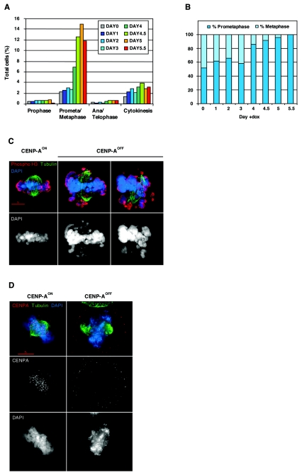
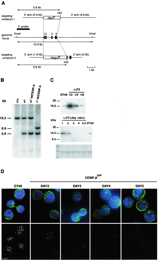
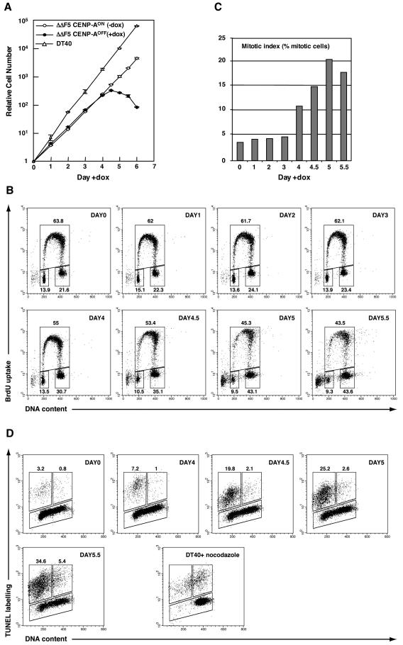
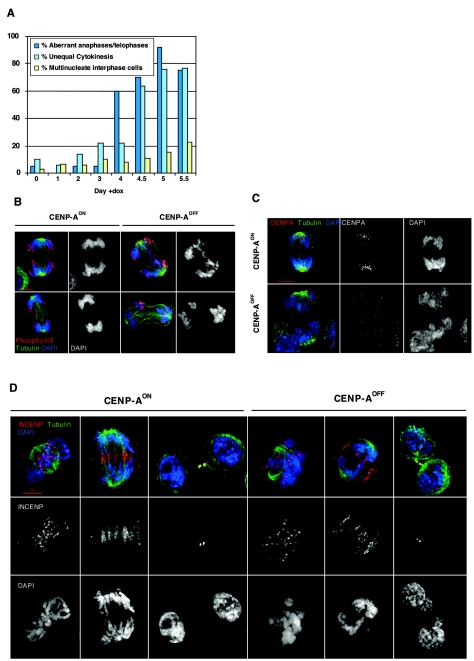
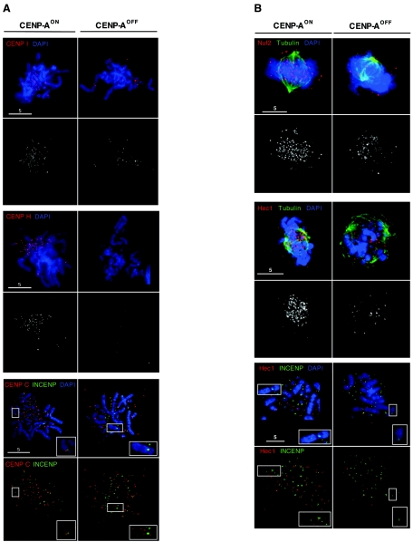
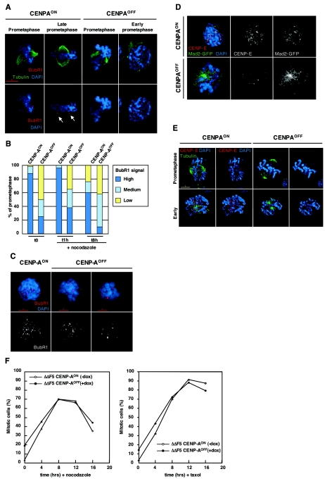
CENP-A is an evolutionarily conserved, centromere-specific variant of histone H3 that is thought to play a central role in directing kinetochore assembly and in centromere function. Here, we have analyzed the consequences of disrupting the CENP-A gene in the chicken DT40 cell line. In CENP-A-depleted cells, kinetochore protein assembly is impaired, as indicated by mislocalization of the inner kinetochore proteins CENP-I, CENP-H, and CENP-C as well as the outer components Nuf2/Hec1, Mad2, and CENP-E. However, BubR1 and the inner centromere protein INCENP are efficiently recruited to kinetochores. Following CENP-A depletion, chromosomes are deficient in proper congression on the mitotic spindle and there is a transient delay in prometaphase. CENP-A-depleted cells further proceed through anaphase and cytokinesis with unequal chromosome segregation, suggesting that some kinetochore function remains following substantial depletion of CENP-A. We furthermore demonstrate that CENP-A-depleted cells exhibit a specific defect in maintaining kinetochore localization of the checkpoint protein BubR1 under conditions of checkpoint activation. Our data thus point to a specific role for CENP-A in assembly of kinetochores competent in the maintenance of mitotic checkpoint signaling.


Figures

FIG. 3.
FIG. 3.
CENP-A-deficient cells are delayed in prometaphase. (A) Distribution of mitotic stages in CENP-AON (day 0) and CENP-AOFF cells during the depletion time course. Counting was performed on cells stained for tubulin, phosphohistone H3, and DNA. Five hundred to 2,000 cells, including at least 100 mitotic cells, were counted for each time point. (B) Ratio of prometaphase and metaphase cells in the CENP-AON (day 0) and CENP-AOFF populations during the depletion time course. Fifty prometaphase/metaphase cells with a bipolar spindle where both poles were localized in the same plane were counted for each time point. Equatorial chromosome alignment defined the metaphase stage. (C) CENP-AON and CENP-AOFF (doxycycline, day 4.5) were stained for tubulin (green), phosphohistone H3 (red), and DNA (blue). Metaphase cells as observed in control CENP-AON cells were rarely observed in CENP-AOFF cells. Instead, the frequency of cells with misaligned chromosomes increased. (D) CENP-AON and CENP-AOFF (doxycycline, day 4.5) were stained for tubulin (green), CENP-A (red), and DNA (blue). Kinetochore signals were no longer observed on CENP-A-deficient prometaphases.
FIG. 1.
FIG. 1.
Generation of a conditional CENP-A−/− clone. (A) Schematic representation of the chicken CENP-A locus and the gene targeting constructs. Black boxes indicate the position of exons. HIII indicates HindIII sites. (B) Southern blot analysis of wild-type (+/+), heterozygous mutant (+/−), heterozygous mutant with random integration of the Tet-repressible CENP-A transgene (+/− tetCENP-A) and homozygous (−/−) mutant clones. DNA was digested with XmaI and HindIII and hybridized with the 5′ probe shown in panel A. First and second targeted events are diagnosed by the appearance of 5.9- and 6.9-kb restriction fragments replacing the cognate 10.3-kb fragment. (C) Western blot analysis of CENP-A expression. Nuclear extracts from DT40 cells or the CENP-A−/− cell line expressing the Tet-repressible transgene (ΔΔF5) were separated on a 15% sodium dodecyl sulfate-polyacrylamide gel electrophoresis gel and analyzed by immunoblotting with affinity-purified rabbit anti-chicken CENP-A. Coomassie blue staining of histone proteins in the extracts was used as a loading control. (Upper panel) Serial dilutions of ΔΔF5 extracts were loaded to quantify overexpression level in the ΔΔF5 cell line. (Lower panel) Time course of CENP-A repression following addition of doxycycline in the ΔΔF5 cell line. (D) Immunofluorescence analysis of DT40 and ΔΔF5 cell line at the indicated time following addition of doxycycline. Cells were stained for CENP-A (red), α-tubulin (green), and DNA (blue). Images for each time point were acquired using the same acquisition parameters. Background signal is increased at late time points (days 4 and 5) due to nonspecific binding of the primary antibody (see main text).
FIG. 2.
FIG. 2.
Cell cycle analysis of CENP-AOFF cells. (A) Growth curve of parental DT40 and ΔΔF5 cells in the presence or absence of doxycycline. (B) Cell cycle distribution of ΔΔF5 cells after addition of doxycycline as measured by bromodeoxyuridine (BrdU) incorporation and DNA content in flow cytometry analysis. Cells were pulse-labeled with BrdU and stained with FITC-anti-BrdU to detect BrdU incorporation (vertical axis, logarithmic scale) and propidium iodide to detect total DNA (horizontal axis, linear scale). The lower-left gate identifies G1 cells, the upper gate identifies cells incorporating BrdU (S phase), and the lower-right gate represents G2/M cells. The numbers show the percentage of cells falling in each gate, excluding the apoptotic sub-G1 cells. (C) Mitotic index of ΔΔF5 cells following addition of doxycycline. Fraction of mitotic cells was determined by flow cytometry analysis of phosphohistone H3 staining. (D) Flow cytometric analysis of apoptosis and cell cycle in the ΔΔF5 cell line at the indicated time following addition of doxycycline. DNA strand breaks were labeled by TUNEL reaction using fluorescein-conjugated dUTP (vertical axis, logarithmic scale), and cells were stained with propidium iodide to determine the DNA content (horizontal axis, linear scale). The upper-left gate identifies positive apoptotic cells with a 2N DNA content, whereas the upper-right gate identifies positive apoptotic cells with a 4N DNA content. DT40 cells treated with nocodazole for 16 h were used as a control for apoptosis occurring with a 4N DNA content.
FIG. 4.
FIG. 4.
CENP-A-deficient cells exhibit severe chromosome segregation defects. (A) Quantification of anaphases/telophases with lagging chromosomes, unequal cytokinesis, or multinucleate interphase cells in CENP-AON (day 0) and CENP-AOFF cells. The percentage shown in each category represents the fraction of aberrant events out of the total number of events falling into the category. For each time point until day 5, the anaphases/telophases, cytokinesis, and interphases recorded were 20, 50, and 500, respectively. For day 5.5, the values were 8, 13, and 500, respectively. (B) CENP-AON and CENP-AOFF (doxycycline, day 4.5) anaphases (upper panel) and telophases (lower panel) were stained for tubulin (green), phosphohistone H3 (red), and DNA (blue). Few or multiple lagging chromosomes were frequently observed in CENP-AOFF cells. (C) CENP-AON and CENP-AOFF (doxycycline, day 4.5) anaphase cells stained for tubulin (green), CENP-A (red), and DNA (blue). Discrete CENP-A signals located at poles in CENP-AON cells were no longer visible in CENP-AOFF cells. (D) CENP-AON and CENP-AOFF (doxycycline, day 4.5) cells in prometaphase, anaphase, or cytokinesis stained for tubulin (green), INCENP (red), and DNA (blue). INCENP localizes at kinetochores in CENP-AOFF prometaphase cells and transfers to the midzone in missegregating CENP-AOFF cell anaphases. Cytokinesis in CENP-AOFF cells resulted in daughter cells with several nuclei of unequal size.
FIG. 5.
FIG. 5.
CENP-AOFF cells are depleted of the inner kinetochore proteins CENP-I, CENP-H, and CENP-C and of the Nuf2/Hec1 complex. (A) CENP-AON and CENP-AOFF metaphase spread (doxycycline, day 5) cells were stained for CENP-I (red, upper panel), CENP-H (red, middle panel), CENP-C (red, lower panel), and DNA (blue). Cells stained with CENP-C were also stained with INCENP (green, lower panel). While CENP-AON cells display strong kinetochore signals for CENP-I, CENP-H, and CENP-C, CENP-AOFF cells showed reduction of signal number and/or intensity for CENP-I and CENP-H. CENP-C signals in CENP-AOFF cells become diffuse and were no longer kinetochore localized on mitotic chromosomes. Inset panels show the inner centromere localization of INCENP and the absence of CENP-C staining for the chromosomeindicated by the box. (B) CENP-AON and CENP-AOFF prometaphase cells (doxycycline, day 5) were stained for tubulin (green), DNA (blue), and Nuf2 (red, upper panel) or Hec1 (red, middle panel). Nuf2 and Hec1 staining is strongly reduced in CENP-AOFF cells. CENP-AON and CENP-AOFF metaphase spreads (lower panel) were stained with Hec1 (Red), INCENP (green), and DNA (blue). Inset panels show that kinetochore localization of Hec1 is abolished in CENP-AOFF cells. For all immunofluorescence stainings, control slides stained with the anti-CENP-A antibody were included and no significant chromosomal signal could be detected in CENP-AOFF cells.
FIG. 6.
FIG. 6.
Localization of the checkpoint proteins BubR1, CENP-E, and Mad2 is affected in CENP-A-depleted cells. (A) CENP-AON and CENP-AOFF cells (doxycycline, day 4.5) were stained for tubulin (green), BubR1 (red), and DNA (blue). Unaligned chromosomes in CENP-AON prometaphase cells have strong BubR1 signals. In late prometaphase, signal intensity decreases upon chromosome alignment while out-of-the-plate chromosomes retain strong staining, as indicated by arrows. CENP-AOFF cells only retain a few kinetochores staining on misaligned chromosomes. CENP-AOFF cells still exhibit strong staining in early prometaphase. (B) Quantitation of CENP-AON and CENP-AOFF (doxycycline, day 4.5) prometaphase cells with high (many kinetochores), medium (few kinetochores), or weak (very few kinetochores) BubR1 signals. Cells were either nontreated (t = 0) or received nocodazole for 1 h or 8 h. Fifty prometaphase cells were counted in each case. (C) CENP-AON and CENP-AOFF (doxycycline, day 4.5) were treated with nocodazole (1 h) and stained for BubR1 (red) and DNA (blue). CENP-AON prometaphase cells retain a strong BubR1 staining when treated with the spindle drug, while most of CENP-AOFF cells had either few (CENP-AOFF, left panel) or very few (CENP-AOFF, right panel) kinetochores stained. (D) Nocodazole-treated (2 h) CENP-AON and CENP-AOFF cells stably expressing a Mad2-GFP construct were stained for CENP-E (red) and DNA (blue). Kinetochore localization of CENP-E and Mad2-GFP was lost in nocodazole-arrested prometaphase cells. (E) CENP-AON and CENP-AOFF cells (doxycycline, day 4.5) were stained for tubulin (green), CENP-E (red), and DNA (blue). CENP-E signals were hardly detectable in prometaphase cells, whereas some signals could be detected in early prometaphase. (F) Mitotic index of CENP-AON and CENP-AOFF (doxycycline day 4.5) during a 16-h time course of nocodazole or paclitaxel treatment. The fraction of mitotic cells was determined by flow cytometry analysis of phosphohistone H3 staining. For all immunofluorescence stainings, control slides stained with the anti-CENP-A antibody were included and no significant chromosomal signal could be detected in CENP-AOFF cells.

References

    1. Adams, R. R., M. Carmena, and W. C. Earnshaw. 2001. Chromosomal passengers and the (aurora) ABCs of mitosis. Trends Cell Biol. 11:49-54. - PubMed
    1. Adams, R. R., H. Maiato, W. C. Earnshaw, and M. Carmena. 2001. Essential roles of Drosophila inner centromere protein (INCENP) and aurora B in histone H3 phosphorylation, metaphase chromosome alignment, kinetochore disjunction, and chromosome segregation. J. Cell Biol. 153:865-880. - PMC - PubMed
    1. Biggins, S., and C. E. Walczak. 2003. Captivating capture: how microtubules attach to kinetochores. Curr. Biol. 13:R449-R460. - PubMed
    1. Black, B. E., D. R. Foltz, S. Chakravarthy, K. Luger, V. L. Woods, Jr., and D. W. Cleveland. 2004. Structural determinants for generating centromeric chromatin. Nature 430:578-582. - PubMed
    1. Blower, M. D., and G. H. Karpen. 2001. The role of Drosophila CID in kinetochore formation, cell-cycle progression and heterochromatin interactions. Nat. Cell Biol. 3:730-739. - PMC - PubMed

Publication types

MeSH terms

LinkOut - more resources