. 2005 May;25(10):4010-22.
doi: 10.1128/MCB.25.10.4010-4022.2005.

The LIM protein Ajuba influences interleukin-1-induced NF-kappaB activation by affecting the assembly and activity of the protein kinase Czeta/p62/TRAF6 signaling complex

Affiliations

The LIM protein Ajuba influences interleukin-1-induced NF-kappaB activation by affecting the assembly and activity of the protein kinase Czeta/p62/TRAF6 signaling complex

Yungfeng Feng et al. Mol Cell Biol. 2005 May.

Abstract

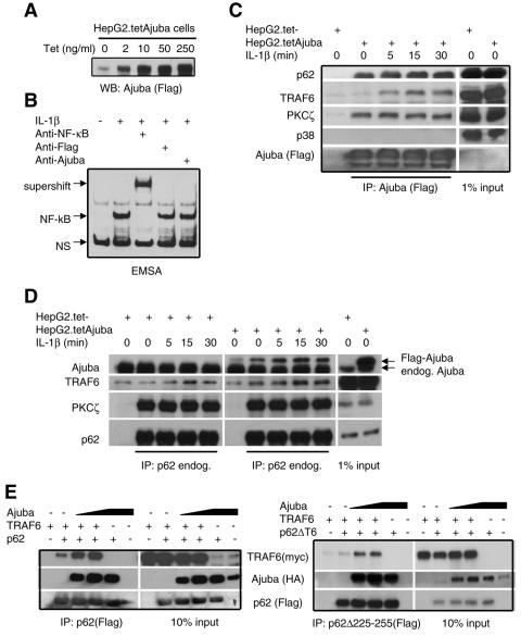
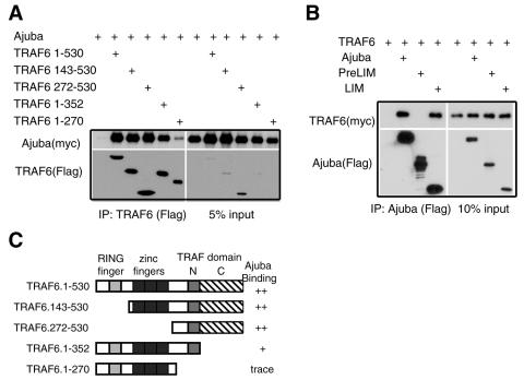
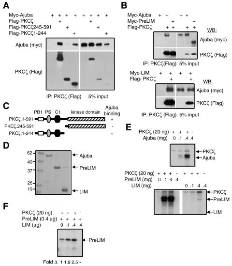
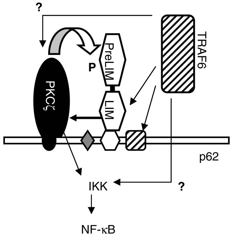
The Zyxin/Ajuba family of cytosolic LIM domain-containing proteins has the potential to shuttle from sites of cell adhesion into the nucleus and thus can be candidate transducers of environmental signals. To understand Ajuba's role in signal transduction pathways, we performed a yeast two-hybrid screen with the LIM domain region of Ajuba. We identified the atypical protein kinase C (aPKC) scaffold protein p62 as an Ajuba binding partner. A prominent function of p62 is the regulation of NF-kappaB activation in response to interleukin-1 (IL-1) and tumor necrosis factor signaling through the formation of an aPKC/p62/TRAF6 multiprotein signaling complex. In addition to p62, we found that Ajuba also interacted with tumor necrosis factor receptor-associated factor 6 (TRAF6) and PKCzeta. Ajuba recruits TRAF6 to p62 and in vitro activates PKCzeta activity and is a substrate of PKCzeta. Ajuba null mouse embryonic fibroblasts (MEFs) and lungs were defective in NF-kappaB activation following IL-1 stimulation, and in lung IKK activity was inhibited. Overexpression of Ajuba in primary MEFs enhances NF-kappaB activity following IL-1 stimulation. We propose that Ajuba is a new cytosolic component of the IL-1 signaling pathway modulating IL-1-induced NF-kappaB activation by influencing the assembly and activity of the aPKC/p62/TRAF6 multiprotein signaling complex.


Figures

FIG. 1.
FIG. 1.
The LIM protein Ajuba associates with the aPKC-interacting protein p62. (A) Stick figure representation of Ajuba. The gray boxes represent the three LIM domains present in the C-terminal LIM region. The PreLIM region is N terminal. (B) Ajuba and p62 associate in human HepG2 cells. Cell extracts were immunoprecipitated with preimmune serum (PI, lane 1) or Ajuba antiserum (lane 2). Bound products were Western blotted for the presence of p62. In lane 3, 0.5% of the amount of cell extract used for immunoprecipitation (input) was run. (C) Ajuba and p62 colocalize in cells. Fibroblasts were transfected with Flag-tagged p62 alone (panel i), myc-tagged Ajuba alone (panel ii), or both p62-Flagand myc-Ajuba (panels iii to v) and immunostained for p62 (anti-Flag, red, panels i and iii) or Ajuba (anti-myc, green, panels ii and iv). Panel v is a merged image of panels iii and iv. Arrows identify vesicular, endosomal structures. Arrowheads identify focal adhesion sites. (D) Ajuba and LIMD1 but not Zyxin or LPP associate with p62. HEK293 cells were transfected with p62 (Flag-tagged) and myc-tagged LIM proteins, as indicated. p62 was immunoprecipitated (anti-Flag), and bound products were Western blotted for the presence of LIM protein (anti-myc, upper panels) and p62 (anti-Flag, lower panels). For each set, input controls were run (5% of amount used for immunoprecipitation). (E) Mapping of the region of Ajuba that interacts with p62. HEK293 cells were transfected with myc-tagged p62 and Flag-tagged isoforms of Ajuba, as indicated. Ajuba was immunoprecipitated (anti-Flag), and bound products were Western blotted for the presence of p62 (anti-myc, left upper panel) and Ajuba isoforms (anti-Flag, left lower panel). Five percent of the cell extract used for immunoprecipitation was run as an input control (right panels).
FIG. 2.
FIG. 2.
Ajuba is required for efficient activation of NF-κB by IL-1 and TNF. (A) Two sets of litter-matched Ajuba−/− and control wt MEFs were stimulated with IL-1β (10 ng/ml) or TNF-α (10 ng/ml) for the indicated times. Nuclei were isolated, and EMSA with NF-κB-specific oligonucleotides was performed, as described in the text. Equal amounts of nuclear extracts were used in each lane. The percent NF-κB activity in Ajuba null cells compared to control wt cells is indicated under each lane. (B) Ajuba−/− and control wt MEFs were treated as in panel A, and cytosolic fractions from the same experimental cells were Western blotted for phospho-IκΒ (upper panel) and total IκΒα protein (lower panel). The percent NF-κB activity in Ajuba null cells compared to control wt cells is indicated under each lane. (C) Ajuba−/− (left lane) and Ajuba null MEFs stably expressing exogenous Ajuba (right lane) were treated as described for panel A, and nuclear EMSA for NF-κB activity was performed. The percent NF-κB activity in Ajuba-rescued cells compared to Ajuba null cells is indicated under each lane. The lower panel is an anti-Ajuba Western blot comparing the level of Ajuba protein in wt (+/+), Ajuba null (−/−), and Flag-tagged Ajuba-rescued Ajuba null MEFs (−/−, +A). Equal amounts of protein were loaded in each lane. (D) Zyxin−/− and control wt MEFs were stimulated with IL-1β (10 ng/ml) for the indicated times. Nuclei were isolated, and EMSA with NF-κB-specific oligonucleotides was performed, as in panel A. Equal amounts of nuclear extracts were used in each lane. The percent NF-κB activity in Zyxin null cells compared to control wt cells is indicated under each lane. (E) Ajuba−/− and control wt MEFs were treated as in panel A, and the same cytosolic fractions were Western blotted for phospho-JNK, total JNK-1, phospho-ERK, total ERK1, phospho-p38, and total p38 protein. Equal amounts of protein were loaded in each lane.
FIG. 3.
FIG. 3.
Ajuba is required for IL-1-induced NF-κB activation in vivo through activation of IKK activity. (A) Ajuba−/− and control wt mice were injected with IL-1β for the indicated times. Nuclear extracts from the lungs of these mice were analyzed by NF-κB EMSA assays (upper panel), nuclear p65 subunit of NF-κB Western blotting (middle panel), and nucleophosmin Western blotting of nuclear extracts as a loading control (lower panel). (B) Semiquantitative RT-PCR analysis of lungs from IL-1β-injected mice for the level of the NF-κB-responsive gene CXCL2 (MIP-2) (44) (upper panel). Loading control is GAPDH (lower panel). (C) IKK in vitro kinase assay of cytosolic extracts from the lungs of the mice described for panel A. GST-IκΒα(1-54) was used as exogenous substrate.
FIG. 4.
FIG. 4.
A unique domain in p62, not overlapping with aPKC or TRAF6 binding sites, directs its interaction with Ajuba. HEK293 cells were cotransfected with myc-tagged Ajuba and Flag-tagged p62. p62 (anti-Flag) was immunoprecipitated, and bound products were Western blotted for the presence of Ajuba (anti-myc) and p62 (anti-Flag) (left panels). Right panels show input controls. (A) Mapping of the p62 truncation mutants. (B and C) Mapping of p62 deletion mutants. (D) Stick figure representation of p62 mutants tested and those that associate with Ajuba. AID is the atypical PKC-interacting domain, ZZ is the zinc finger RIP-interacting domain, LB is the novel LIM protein binding domain, and T6 is the TRAF6 binding domain.
FIG. 5.
FIG. 5.
Ajuba recruits TRAF6 into the p62/PKCζ signaling complex. (A) HepG2 cells containing tet-regulated Ajuba (Flag-tagged) were stimulated with differing concentrations of tetracycline, and Ajuba (anti-Flag) Western blotting was performed. (B) NF-κB supershift assay. HepG2 cells containing tet-regulated Ajuba (Flag-tagged) were induced with tetracycline (2 ng/ml) and then stimulated with IL-1β (10 ng/ml). Nuclei were isolated, and NF-κB EMSA was performed in the absence (lane 2) or presence of antibodies to the p50 subunit of NF-κB (lane 3), Flag (lane 4), or Ajuba (lane 5). NS indicates nonspecific bands. (C and D) HepG2 cells containing tet-regulated Ajuba (Flag-tagged) were induced with tetracycline (2 ng/ml) and then stimulated with IL-1β (10 ng/ml) for the indicated times. Ajuba (anti-Flag) (C) or endogenous p62 (D) were immunoprecipitated, and bound products were Western blotted for the presence of endogenous p62, TRAF6, PKCζ, and p38 as a negative control. Right panels show input controls. (E) HEK293 cells were cotransfected with fixed amounts of TRAF6 (myc-tagged), either p62 (Flag-tagged) (left panels) or p62.Δ225-255 (right panels), and increasing amounts of HA-tagged Ajuba. p62 was immunoprecipitated (anti-Flag), and bound products were Western blotted for the presence of TRAF6 (anti-myc), Ajuba (anti-HA), and p62 (anti-Flag) (left panels). Right panels show input controls.
FIG. 6.
FIG. 6.
Ajuba interacts with TRAF6. (A) Mapping of the TRAF6 domains required for an interaction with Ajuba. HEK293 cells were cotransfected with myc-tagged Ajuba and Flag-tagged TRAF6 isoforms. TRAF6 (anti-Flag) was immunoprecipitated, and bound products were Western blotted for the presence of Ajuba (anti-myc) and TRAF6 (anti-Flag) (left panels). Right panels show input controls. (B) Mapping of the Ajuba regions required for an interaction with TRAF6. HEK293 cells were cotransfected with Flag-tagged Ajuba isoforms and myc-tagged TRAF6. Ajuba isoforms (anti-Flag) were immunoprecipitated, and bound products were Western blotted for the presence of TRAF6 (anti-myc) and Ajuba (anti-Flag) (left panels). Right panels show input controls. (C) Stick figure representation of TRAF6 mutants and their association with Ajuba. The light gray box is the RING finger domain, the black box is the five zinc fingers, the dark gray box is the N-terminal region of the TRAF domain, and the hatched box is the C-terminal region of the TRAF domain.
FIG. 7.
FIG. 7.
Ajuba interacts with PKCζ, activates PKCζ, and is a substrate of PKCζ. (A) Mapping of the PKCζ region required to interact with Ajuba. HEK293 cells were cotransfected with myc-tagged Ajuba and Flag-tagged PKCζ mutants. PKCζ isoforms were immunoprecipitated (anti-Flag), and bound products were Western blotted for the presence of Ajuba (anti-myc) (left panels). Right panels show input controls. (B) Mapping of the Ajuba region required to interact with PKCζ, as described for panel A. (C) Stick figure representation of PKCζ mutants and their interaction with Ajuba. (D) Coomassie-stained gel of baculovirus-produced and purified Ajuba, PreLIM peptide of Ajuba, and LIM peptide of Ajuba. Molecular weight markers in thousands are on the left. (E) In vitro PKCζ kinase assays. A fixed amount of purified PKCζ (20 ng) was mixed with increasing amounts of Ajuba protein (upper panel) or PreLIM and LIM peptides (lower panel). Arrows identify the location of PKCζ and Ajuba peptides. (F) Purified PKCζ (20 ng) was mixed with PreLIM peptide (400 ng). Increasing amounts of Ajuba LIM peptide were added, and an in vitro kinase reaction was performed. In this assay, more nonradioactive ATP was present than in panel E. The arrow identifies a phosphorylated PreLIM peptide. The increase (n-fold) in PreLIM phosphorylation relative to that observed in the absence of LIM peptide (set at 1) is shown below each lane.
FIG. 8.
FIG. 8.
Working model for how Ajuba influences IL-1-induced NF-κB activation. PKCζ and the PKCζ binding site in p62 (AID) are in black. Ajuba and the LIM protein binding site in p62 (LB) are in white. TRAF6 and the TRAF6 binding site in p62 (T6) are hatched. The LIM region of Ajuba interacts with both TRAF6 and PKC. In vitro, the LIM region of Ajuba activates PKC, and the PreLIM region of Ajuba can be phosphorylated by PKC. Whether phosphorylation of Ajuba regulates either the assembly of the multiprotein complex or the capacity of the complex to activate IKK is not known.

Similar articles

Cited by

References

    1. Arch, R. H., R. W. Gedrich, and C. B. Thompson. 1998. Tumor necrosis factor receptor-associated factors (TRAFs)—a family of adapter proteins that regulates life and death. Genes Dev. 12:2821-2830. - PubMed
    1. Bach, I. 2000. The LIM domain: regulation by association. Mech. Dev. 91:5-17. - PubMed
    1. Baud, V., Z. G. Liu, B. Bennett, N. Suzuki, Y. Xia, and M. Karin. 1999. Signaling by proinflammatory cytokines: oligomerization of TRAF2 and TRAF6 is sufficient for JNK and IKK activation and target gene induction via an amino-terminal effector domain. Genes Dev. 13:1297-1308. - PMC - PubMed
    1. Burstein, E., and C. S. Duckett. 2003. Dying for NF-κB? Control of cell death by transcriptional regulation of the apoptotic machinery. Curr. Opin. Cell Biol. 15:732-737. - PubMed
    1. Crawford, A. W., and M. C. Beckerle. 1991. Purification and characterization of zyxin, an 82,000-dalton component of adherens junctions. J. Biol. Chem. 266:5847-5853. - PubMed

Publication types

MeSH terms

Substances