. 2005 May;25(10):4092-104.
doi: 10.1128/MCB.25.10.4092-4104.2005.

The nuclear import of TAF10 is regulated by one of its three histone fold domain-containing interaction partners

Affiliations

The nuclear import of TAF10 is regulated by one of its three histone fold domain-containing interaction partners

Evi Soutoglou et al. Mol Cell Biol. 2005 May.

Abstract

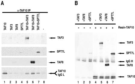
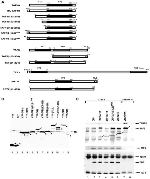
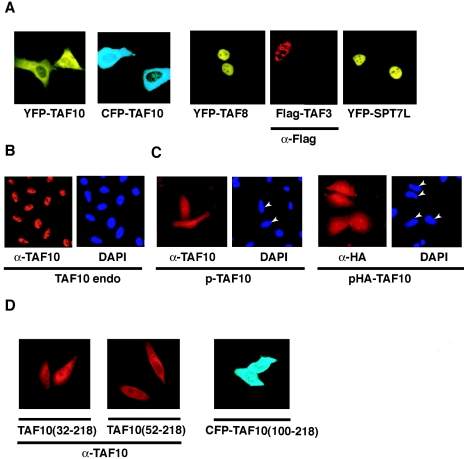
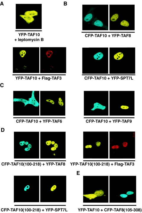
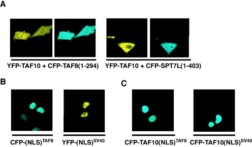
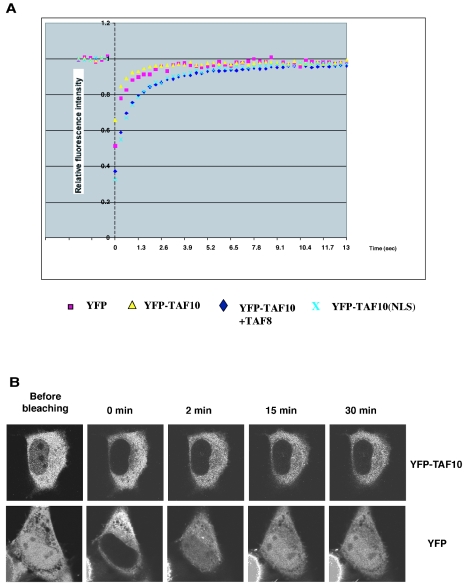
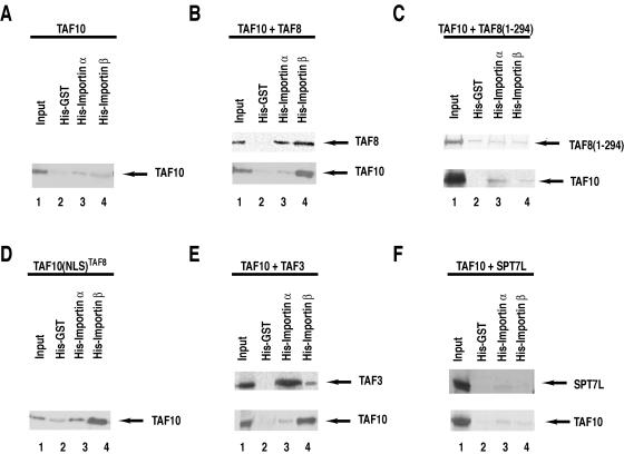
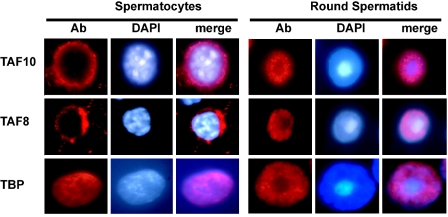
TFIID, comprising the TATA box binding protein (TBP) and 13 TBP-associated factors (TAFs), plays a role in nucleation in the assembly of the RNA polymerase II preinitiation complexes on protein-encoding genes. TAFs are shared among other transcription regulatory complexes (e.g., SAGA, TBP-free TAF-containing complex [TFTC], STAGA, and PCAF/GCN5). Human TAF10, a subunit of both TFIID and TFTC, has three histone fold-containing interaction partners: TAF3, TAF8, and SPT7Like (SPT7L). In human cells, exogenously expressed TAF10 remains rather cytoplasmic and leptomycin B does not affect this localization. By using fluorescent fusion proteins, we show that TAF10 does not have an intrinsic nuclear localization signal (NLS) and needs one of its three interaction partners to be transported into the nucleus. When the NLS sequences of either TAF8 or SPT7L are mutated, TAF10 remains cytoplasmic, but a heterologous NLS can drive TAF10 into the nucleus. Experiments using fluorescence recovery after photobleaching show that TAF10 does not associate with any cytoplasmic partner but that once transported into the nucleus it binds to nuclear structures. TAF10 binding to importin beta in vitro is dependent on the coexpression of either TAF8 or TAF3, but not SPT7L. The cytoplasmic-nuclear transport of TAF10 is naturally observed during the differentiation of adult male germ cells. Thus, here we describe a novel role of the three mammalian interacting partners in the nuclear localization of TAF10, and our data suggest that a complex network of regulated cytoplasmic associations may exist among these factors and that this network is important for the composition of different TFIID and TFTC-type complexes in the nucleus.


Figures

FIG. 1.
FIG. 1.
Human TAF10 interacts with TAF3, TAF8, and SPT7L in vitro. (A) TAF3, TAF8, TAF10, and SPT7L were expressed individually or TAF10 was coexpressed with one of its potential partners (as indicated) in Sf9 cells by using the baculovirus expression system. Forty-eight hours after infection, WCEs were made, TAF10-associated proteins were subjected to IP with an anti-TAF10 mAb, the beads were extensively washed, boiled, and loaded onto an SDS-polyacrylamide gel, and TAF10-associated proteins were analyzed by Western blotting using the indicated antibodies. (B) TAF10 was first bound to protein G-Sepharose-anti-TAF10 antibody beads, the beads were extensively washed, and then protein extracts containing one of the three partners were incubated (as indicated) with the TAF10-containing beads (lanes 5 to 7) or with protein G-Sepharose antibody beads alone as controls (lanes 1 to 3) for 1 h at room temperature, and beads were extensively washed and bound proteins analyzed as described for panel A. IgG, immunoglobulin G (L, light chain; H, heavy chain).
FIG. 2.
FIG. 2.
(A) Schematic representation of the TAF10-, TAF8-, TAF3-, and SPT7L-containing fusion proteins used in the HeLa cell transfection experiments. Most of the constructs were generated as CFP and/or YFP fusion proteins. The histone fold domain (HFD), the proline-rich domain of TAF8 (TAPD), the PHD finger, and the NLSs of the factors are indicated. The numbers refer to amino acid positions in each protein. N-ter, N terminus. (B) The YFP- and CFP-containing expression vectors express the correct fusion proteins in transfected HeLa cells. HeLa cells were transfected (as indicated), WCEs prepared, and 50 μg protein separated by SDS-PAGE and analyzed by Western blotting using an anti-GFP antibody. In each lane, the specifically expressed protein is labeled with an arrowhead. NS, nonspecific protein also present in nontransfected cell extracts. (C) The expressed YFP and CFP fusion transcription factors are functional since they integrate into their respective complexes. From the indicated extracts analyzed as described for panel B, either anti-TBP (lanes 1 to 6) or anti-TRRAP (lanes 7 to 8) IP was carried out, and bound proteins were analyzed as described in the legend to Fig. 1. −, antibody control without extract. The CFP or YFP fusion proteins incorporated into either a TBP- or a TRRAP-containing complex are labeled with arrowheads. Western blotting with anti-TRRAP, anti-TAF5, anti-TAF6, and anti-TBP was carried out to verify that the anti-TBP (α-TBP) and anti-TRRAP IPs worked as expected. IgG, immunoglobulin G (L, light chain; H, heavy chain).
FIG. 3.
FIG. 3.
Exogenously expressed TAF10 localizes mostly to the cytoplasm of the cells, and TAF3, TAF8, and SPT7L are homogenously distributed within the nucleus but excluded from the nucleolus. (A to D) HeLa cells were either nontransfected (B) or transfected with the indicated expression vectors, and the expressed proteins were visualized by fluorescence microscopy either directly or by using the indicated primary mouse antibodies and a Cy3-labeled secondary antibody. Where indicated, the nuclei of the cells were visualized with DAPI staining. In panel C, the nuclei of the transfected cells are indicated with arrowheads. If antibodies (α) were used to visualize the proteins, they are indicated under the corresponding photos. TAF10 endo, endogenous TAF10.
FIG. 4.
FIG. 4.
Exogenously expressed TAF10 becomes exclusively nuclear when cells are cotransfected with one of TAF10's HF domain-containing interaction partners. The HF domain of TAF10 and the partner are required for nuclear localization. (A to E) HeLa cells were cotransfected with the indicated CFP- and YFP-containing expression vectors, and localization of the expressed proteins was visualized by fluorescence microscopy. Flag-TAF3 was visualized by immunofluorescence. In panel A, pYFP-TAF10-transfected cells were incubated for 4 h with 20 ng/ml leptomycin B. The images shown in each panel are representative of all the transfected cells.
FIG. 5.
FIG. 5.
The nuclear localization of exogenously expressed TAF10 depends on the presence of an NLS either located in the interaction partner itself or fused directly to TAF10. (A to C) HeLa cells were cotransfected with the indicated CFP- and YFP-containing expression vectors, and localization of the expressed proteins was visualized by fluorescence microscopy. The images shown in each panel are representative of all the transfected cells.
FIG. 6.
FIG. 6.
YFP-TAF10 diffuses freely within the cytoplasm but binds to structures in the nucleus. (A) Quantitative FRAP analysis of YFP, YFP-TAF10 in the cytoplasm, nuclear YFP-TAF10-NLSTAF8, and nuclear YFP-TAF10 after coexpression with TAF8. YFP and cytoplasmic YFP-TAF10 show similar, very fast recovery kinetics in the cytoplasm (250 ms), whereas nuclear YFP-TAF10 shows much slower recovery kinetics (3 s) than its cytoplasmic counterpart. (B) Confocal series of pictures of transfected cells before and after photobleaching of the nuclear fluorescent pool of either YFP or YFP-TAF10. YFP shows fast and almost complete recovery within the first 5 min after bleaching. YFP-TAF10 exhibits only minimal recovery even after 30 min.
FIG. 7.
FIG. 7.
In vitro TAF10 binding to importin β is dependent on the coexpression of either TAF8 or TAF3, but not SPT7L. (A to F) Six-His-tagged GST, six-His-tagged importin α, and six-His-tagged importin β were expressed in Escherichia coli and bound to Ni-NTA beads that were then extensively washed. HeLa cells were cotransfected with CFP- and YFP- or Flag-containing expression vectors (as indicated at the top of each panel); 48 h after transfection, WCEs were prepared and the extracts were incubated with the control His-tagged GST- as well as the importin α- and importin β-containing beads. Beads were then washed and bound proteins analyzed by Western blotting with an anti-GFP antibody. A small amount (0.1%) of the input extract was also analyzed on the same blots.
FIG. 8.
FIG. 8.
Endogenous TAF10 and TAF8 are cytoplasmic in primary adult spermatocytes but not in round spermatids. Squash preparations from segments of defined stages of the mouse adult seminiferous tubuli were obtained by transillumination-assisted microdissection and probed with anti-TAF10, anti-TAF8, or anti-TBP antibody (left panels of each series). The middle and right panels show the corresponding DAPI-stained DNA and merged images, respectively. Magnification, ×100. Ab, antibody.

References

    1. Albright, S. R., and R. Tjian. 2000. TAFs revisited: more data reveal new twists and confirm old ideas. Gene 242:1-13. - PubMed
    1. Bell, B., E. Scheer, and L. Tora. 2001. Identification of hTAF(II)80 delta links apoptotic signaling pathways to transcription factor TFIID function. Mol. Cell 8:591-600. - PubMed
    1. Bell, B., and L. Tora. 1999. Regulation of gene expression by multiple forms of TFIID and other novel TAFII-containing complexes. Exp. Cell Res. 246:11-19. - PubMed
    1. Berthelsen, J., C. Kilstrup-Nielsen, F. Blasi, F. Mavilio, and V. Zappavigna. 1999. The subcellular localization of PBX1 and EXD proteins depends on nuclear import and export signals and is modulated by association with PREP1 and HTH. Genes Dev. 13:946-953. - PMC - PubMed
    1. Brand, M., C. Leurent, V. Mallouh, L. Tora, and P. Schultz. 1999. Three-dimensional structures of the TAFII-containing complexes TFIID and TFTC. Science 286:2151-2153. - PubMed

Publication types

MeSH terms