. 2005 May;25(10):4200-10.
doi: 10.1128/MCB.25.10.4200-4210.2005.

Expression, regulation, and requirement of the toll transmembrane protein during dorsal vessel formation in Drosophila melanogaster

Affiliations

Expression, regulation, and requirement of the toll transmembrane protein during dorsal vessel formation in Drosophila melanogaster

Jianbo Wang et al. Mol Cell Biol. 2005 May.

Abstract

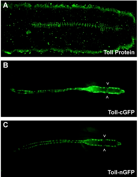
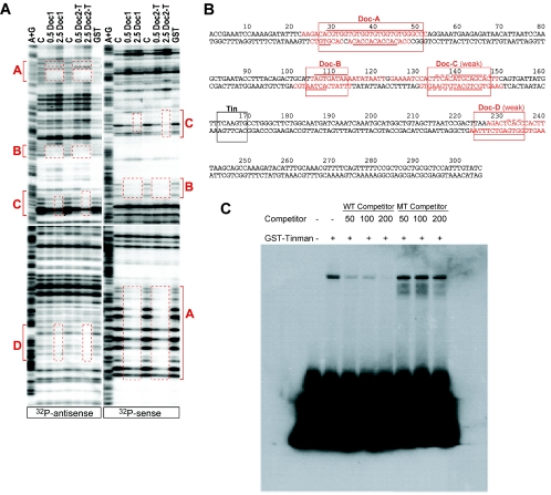
Early heart development in Drosophila and vertebrates involves the specification of cardiac precursor cells within paired progenitor fields, followed by their movement into a linear heart tube structure. The latter process requires coordinated cell interactions, migration, and differentiation as the primitive heart develops toward status as a functional organ. In the Drosophila embryo, cardioblasts emerge from bilateral dorsal mesoderm primordia, followed by alignment as rows of cells that meet at the midline and morph into a dorsal vessel. Genes that function in coordinating cardioblast organization, migration, and assembly are integral to heart development, and their encoded proteins need to be understood as to their roles in this vital morphogenetic process. Here we prove the Toll transmembrane protein is expressed in a secondary phase of heart formation, at lateral cardioblast surfaces as they align, migrate to the midline, and form the linear tube. The Toll dorsal vessel enhancer has been characterized, with its activity controlled by Dorsocross and Tinman transcription factors. Consistent with the observed protein expression pattern, phenotype analyses demonstrate Toll function is essential for normal dorsal vessel formation. Such findings implicate Toll as a critical cell adhesion molecule in the alignment and migration of cardioblasts during dorsal vessel morphogenesis.


Figures

FIG. 1.
FIG. 1.
Toll mRNA and protein expression in cardioblasts of the forming and mature dorsal vessel. (A and D) Embryos at stage 14; (B and E) embryos at stage 15; (C and F) embryos at stage 16. Abbreviations: a, aorta region of the dorsal vessel; cb, cardioblasts; h, heart region of the dorsal vessel.
FIG. 2.
FIG. 2.
Mapping the location of the Toll dorsal vessel transcriptional enhancer. A condensed organization of the Toll gene is shown at the top. The gene is present at position 97D2-3 of the D. melanogaster cytogenetic map. Various DNA regions located upstream of the Toll transcription start site were tested for enhancer function in dorsal vessel (DV) cardioblasts. DNAs possessing enhancer activity were scored as positive (+), while those lacking activity were scored as negative (−). At least four independently derived transgenic lines were assayed for each of the DNAs tested.
FIG. 3.
FIG. 3.
The Toll 305-bp enhancer functions in all cardioblasts of the dorsal vessel. (A) Toll protein expression at lateral surfaces of all cardioblasts in the formed dorsal vessel at embryonic stage 16. Enhancer activity was detected through the analysis of stage 16 embryos from transgenic strains expressing the Toll DNA fused to marker genes encoding cytoplasmic green fluorescent protein (Toll-cGFP) (B) or nuclear green fluorescent protein (Toll-nGFP) (C). Open arrowheads indicate a pair of valvelike ostia in panel B and small nuclei present in Svp/Doc cells in panel C.
FIG. 4.
FIG. 4.
Functional dissection of the Toll 305-bp dorsal vessel enhancer. (Left) The Toll 305-bp DNA is schematized at the top, with Doc binding sites indicated by boxes (Doc-A through Doc-D) and the Tin binding site highlighted as an oval. 5′-truncated and/or site-mutated versions of the Toll enhancer are schematized below the wild-type DNA, with constructs named according to their size in nucleotides and the included mutation. For example, Toll 258 represents a DNA lacking 47 bp of the 5′ sequence, including most of the Doc-A binding region; Toll 258 mTin is the same DNA with a mutated Tin binding site. Activities of the various DNAs in Tin or Svp/Doc subsets of cardioblasts are indicated at the right as positive (+), negative (−), or positive but variable (+/−) in enhancer function. At least four independently derived transgenic lines were assayed for each of the DNAs tested. (Right) Expression patterns of Toll enhancer DNAs in transgenic animals. Stage 16 embryos were assayed for enhancer activity using cytoplasmic β-galactosidase (clacZ) and cGFP markers. (A) Toll 305-clacZ expression in all cardioblasts. (B) Mutation of the Tin-binding site in Toll 305 mTin results in variable and irregular activity of the enhancer. Arrows point out the lack of reporter expression in Tin cardioblasts. (C) Toll 258-clacZ expression solely in Tin cardioblasts. Open arrowheads highlight enhancer inactivity in Svp/Doc cells. (D) Mutation of the Tin-binding site in Toll 258 mTin results in enhancer nonfunction in Tin cardioblasts. (E) Toll 264-cGFP expression solely in Tin cardioblasts. Open arrowheads indicate enhancer inactivity in Svp/Doc cells. (F, G, and H) Toll 270, Toll 276, and Toll 282 DNAs drive cGFP expression strongly in Tin cardioblasts and variably in Svp/Doc cardioblasts. Solid arrowheads point out the variable enhancer function in Svp/Doc cells. (I) Toll 287-cGFP expression in all cardioblasts of the dorsal vessel.
FIG. 5.
FIG. 5.
DNA binding studies on the Toll dorsal vessel enhancer. (A) In vitro binding of Doc proteins to the Toll dorsal vessel enhancer as determined by DNase I footprint analysis. The antisense (left side) and sense (right side) strands of the 305-bp DNA were 32P labeled and incubated with either 0.5 or 2.5 μg of recombinant GST-Doc fusion protein (full-length Doc1 or T-box fragment of Doc2), 5 μg of GST control protein, or control buffer only. Four areas were found to be protected by recombinant Doc proteins (red boxes labeled A, B, C, and D). Sites A and B show significant DNase I protection at both low and high concentrations of Doc proteins; the C and D sites require the higher protein concentration for protection. Site D is poorly protected on the sense strand (not shown). Abbreviations: A+G, adenosine/guanosine ladder produced via Maxam-Gilbert sequencing; C, control buffer; Doc2-T, T-box fragment of Doc2. (B) Sequence of the Toll 305-bp dorsal vessel enhancer. Binding sites of two relevant cardiac transcription factors are indicated by boxes. Tin: demonstrated Tin binding site matching the known Tin consensus element. Doc-A through Doc-D: Doc binding sites corresponding to areas protected in DNase I footprint assays. The Doc-A and Doc-B regions are bound with higher affinity than the Doc-C and Doc-D sites. Lines above and below the enhancer DNA indicate the protected sequences for the sense and antisense strands, respectively. Dashed lines denote additional protection observed only at high concentrations of Doc2-T protein. Red and nonunderlined nucleotides adjacent to protected sites indicate the range between the outermost band affected by footprinting and the next unaffected band that is detectable in the assay. (C) Electrophoretic mobility shift assay demonstrates that Tin selectively binds to a consensus recognition element present in the Toll 305-bp enhancer. GST-Tin fusion protein was used in the binding assay with a 32P-end-labeled double-stranded oligonucleotide that included the Tin binding sequence present at coordinates 163 to 169 of the enhancer DNA. Specificity of Tin binding to the probe was tested by competition with wild-type (WT) and Tin site-mutated (MT) double-stranded oligonucleotides, used at increasing (50, 100, or 200) molar excess concentrations.
FIG. 6.
FIG. 6.
Forced-expression studies of tin and Doc2 with Toll 305-clacZ in the Drosophila mesoderm. (A, E, and I) Noninduced control embryos at stage 11 or 14. Panels A and I show lateral views of embryos; panel E shows a ventral embryo view. (B, F, and J) twi-GAL4;UAS-tin embryos at the indicated stages. Panels B and J show lateral views; panel F is a ventral view. (C, G, and K) twi-GAL4; UAS-Doc2 embryos at the indicated stages. Panels C and K show lateral views; panel G is a ventral view. (D, H, and L) twi-GAL4;UAS-tin;UAS-Doc2 embryos at the indicated stages. Panels D and L show lateral views; panel H is a ventral view. Green indicates β-galactosidase expression under the control of the Toll 305 enhancer, while red indicates D-MEF2 expression in the mesoderm. Arrows point to areas of induced expression of the β-galactosidase marker in CNS midline cells and throughout the mesoderm. (Bottom) Analysis of Toll enhancer function in amnioserosa cells expressing Toll 305-cGFP (M), Toll 258-cGFP (N), or Toll 287-cGFP (O).
FIG. 7.
FIG. 7.
Dorsal vessel phenotypes resultant from mutations in the Toll gene. (A) Toll 305-cGFP expression in a stage 16 wild-type (WT) embryo. (B and C) Toll 305-cGFP expression in two different Tlr3/Tlr4 embryos at stage 16, developed at a nonpermissive temperature. (D) D-MEF2 expression in a stage 15 wild-type embryo. (E and F) D-MEF2 expression in two different Tlr3/Tlr4 embryos at stage 15, developed at a nonpermissive temperature. Open arrowheads emphasize cellular gaps in the dorsal vessel, while filled arrowheads or lines highlight the abnormal positioning of cardioblasts in Toll mutant embryos.

References

    1. Alvarez, A. D., W. Shi, B. A. Wilson, and J. B. Skeath. 2003. pannier and pointedP2 act sequentially to regulate Drosophila heart development. Development 130:3015-3026. - PubMed
    1. Anderson, K. V., G. Jurgens, and C. Nusslein-Volhard. 1985. Establishment of dorsal-ventral polarity in the Drosophila embryo: Genetic studies on the role of the Toll gene product. Cell 42:779-789. - PubMed
    1. Azpiazu, N., and M. Frasch. 1993. tinman and bagpipe: two homeo box genes that determine cell fates in the dorsal mesoderm of Drosophila. Genes Dev. 7:1325-1340. - PubMed
    1. Barolo, S., L. A. Carver, and J. W. Posakony. 2000. GFP and β-Galactosidase transformation vectors for promoter/enhancer analysis in Drosophila. BioTechniques 29:726-732. - PubMed
    1. Bodmer, R. 1993. The gene tinman is required for specification of the heart and visceral muscles in Drosophila. Development 118:719-729. - PubMed

Publication types

MeSH terms

LinkOut - more resources