. 2005 May;25(10):4237-49.
doi: 10.1128/MCB.25.10.4237-4249.2005.

The S1P2 receptor negatively regulates platelet-derived growth factor-induced motility and proliferation

Affiliations

The S1P2 receptor negatively regulates platelet-derived growth factor-induced motility and proliferation

Sravan K Goparaju et al. Mol Cell Biol. 2005 May.

Abstract

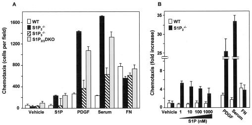
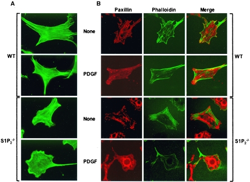
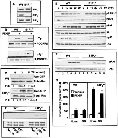
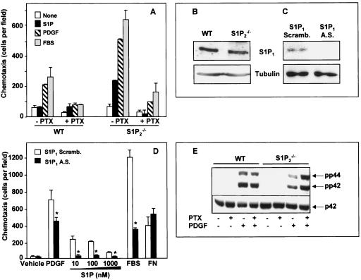
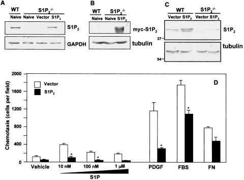
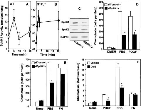
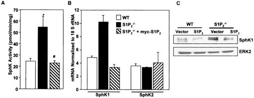
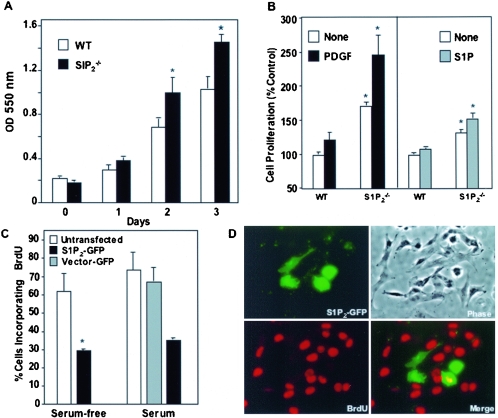
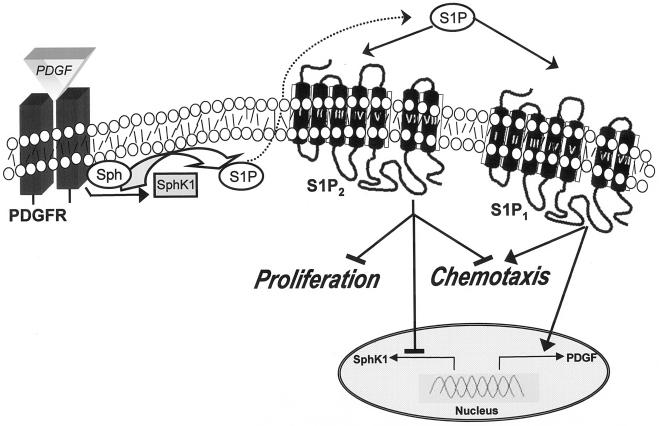
Sphingosine-1-phosphate (S1P), a bioactive sphingolipid metabolite, is the ligand for five specific G protein-coupled receptors, named S1P(1) to S1P(5). In this study, we found that cross-communication between platelet-derived growth factor receptor and S1P(2) serves as a negative damper of PDGF functions. Deletion of the S1P(2) receptor dramatically increased migration of mouse embryonic fibroblasts toward S1P, serum, and PDGF but not fibronectin. This enhanced migration was dependent on expression of S1P(1) and sphingosine kinase 1 (SphK1), the enzyme that produces S1P, as revealed by downregulation of their expression with antisense RNA and small interfering RNA, respectively. Although S1P(2) deletion had no significant effect on tyrosine phosphorylation of the PDGF receptors or activation of extracellular signal-regulated kinase 1/2 or Akt induced by PDGF, it reduced sustained PDGF-dependent p38 phosphorylation and markedly enhanced Rac activation. Surprisingly, S1P(2)-null cells not only exhibited enhanced proliferation but also markedly increased SphK1 expression and activity. Conversely, reintroduction of S1P(2) reduced DNA synthesis and expression of SphK1. Thus, S1P(2) serves as a negative regulator of PDGF-induced migration and proliferation as well as SphK1 expression. Our results suggest that a complex interplay between PDGFR and S1P receptors determines their functions.


Figures

FIG. 1.
FIG. 1.
Deletion of S1P2 increases chemotaxis toward S1P and growth factors. (A) Primary cultures of wild-type (WT), S1P2, S1P3, and single- and S1P2/3 double-knockout (DKO) MEFs were serum starved overnight and then allowed to migrate toward vehicle, S1P (10 nM), PDGF (20 ng/ml), serum (20%), or fibronectin (FN; 20 μg/ml) for 4 h. (B) Cultures of immortalized wild-type and S1P2 knockout MEFs were serum starved overnight and then allowed to migrate for 4 h toward vehicle, S1P, PDGF (20 ng/ml), serum (20%), or fibronectin (20 μg/ml). The data are expressed as fold increase and are means ± SD of triplicate determinations. Migrations of control wild-type and S1P2 knockout MEFs were 26 ± 4 and 29 ± 10 cells per field, respectively. Similar results were obtained in three independent experiments.
FIG. 2.
FIG. 2.
Changes in cytoskeletal architecture and focal complexes of S1P2-deleted MEFs induced by PDGF. Wild-type (WT) and S1P2−/− MEFs were grown on coverslips for 24 h in serum-free medium and then stimulated without (None) or with 20 ng/ml PDGF-BB for 30 min. Cells were then fixed, permeabilized, and stained with (A and B) Cy2-labeled phalloidin to detect actin fibers (green), and (B) adhesion complexes were visualized by confocal microscopy with antipaxillin antibody and Texas red-conjugated secondary antibody. Representative fields of more than 50 cells examined are shown.
FIG. 3.
FIG. 3.
Effect of deletion of S1P2 on PDGF-induced signaling pathways. (A) Expression of S1P1, S1P2, and S1P3 was determined by RT-PCR in wild-type (WT) and S1P2-null MEFs. No PCR products were observed in the absence of RT. (B) Wild-type and S1P2-null MEFs were serum starved overnight, treated with PDGF (20 ng/ml) for 5 min, and lysed, and equal amounts of proteins were immunoprecipitated with anti-PDGRβ (upper panel) or anti-PDGFRα (lower panel) and analyzed by Western blotting with antiphosphotyrosine antibody. Blots were stripped and reprobed with anti-PDGRβ or anti-PDGFRα to demonstrate equal loading. Arrows indicate a PDGFRβ 190-kDa band (upper panel) or a PDGFRα doublet of 150/170 kDa (lower panel). (C) Serum-starved wild-type and S1P2-null MEFs were treated with PDGF for the indicated times, and active Rac was specifically pulled down from cell lysates containing equal amounts of proteins with immobilized recombinant PAK-CRIB and analyzed by Western blotting using anti-Rac antibody. Relative activated levels were normalized to total Rac, and the fold stimulation ± SD from three independent experiments is indicated. Representative results are shown. (D) Serum-starved wild-type and S1P2-null MEFs were treated with vehicle, PDGF, or S1P for 5 min, and activation of PAK1 was determined by Western blot analysis with phospho-PAK1 (Thr423) antibodies (top panel). Equal loading was confirmed by reprobing with antibodies against PAK1, which also cross-react with PAK2 (bottom panel). (E) Wild-type and S1P2-null MEFs were treated with PDGF for the indicated times, and activation of ERK1/2, Akt, and p38 was determined by Western blotting analysis with phospho-specific antibodies. Equal loading was confirmed by reprobing with antibodies against ERK2, Akt, and p38, respectively. Similar results were obtained in three additional experiments. (F) Serum-starved wild-type and S1P2-null MEFs were treated without (lanes 1 and 4) or with SB202190 (10 μM, lanes 2 and 5) or SB202190 (25 μM, lanes 3 and 6) for 15 min and then stimulated with vehicle (lanes 1 to 3) or 20 ng/ml PDGF (lanes 4 to 6) for 10 min. Equal amounts of cell lysate proteins were separated by SDS-PAGE and probed with phospho-specific Akt S473 antibody. Blots were stripped and reprobed with anti-Akt antibody to show equal loading. (G) Serum-starved wild-type and S1P2-null MEFs were treated without or with 10 μM SB202190 (SB) and then allowed to migrate toward vehicle (open bars) or 20 ng/ml PDGF (filled bars) for 4 h.
FIG. 4.
FIG. 4.
Involvement of S1P1 in PDGF-induced chemotaxis. (A) Wild-type (WT) and S1P2−/− MEFs were cultured in serum-free medium for 12 h in the absence or presence of PTX (200 ng/ml). Cells were allowed to migrate for 4 h toward vehicle, S1P (10 nM), PDGF (20 ng/ml), or FBS (20%), and chemotaxis was measured. (B) Expression of S1P1 receptor in wild-type and S1P2−/− MEFs was determined by Western blotting with an antibody against S1P1. The membranes were stripped and reprobed with an antibody against tubulin to show equal loading. (C and D) S1P2−/− cells were transfected with either scrambled (Scramb.; open bars) or antisense (A.S., filled bars) oligonucleotides against S1P1 receptor and (C) analyzed for the expression of S1P1 receptor by Western blotting. Tubulin expression was used as a loading control. (D) Duplicate cultures were serum starved for 12 h and then allowed to migrate toward PDGF (20 ng/ml), serum (20%), or the indicated concentrations of S1P or fibronectin (20 μg/ml). Similar results were obtained in two additional experiments. *, P < 0.05. (E) Wild-type and S1P2−/− MEFs were serum starved overnight in the absence or presence of PTX (100 ng/ml) and then stimulated without or with PDGF (20 ng/ml) for 5 min as indicated. Equal amounts of cell lysate proteins were analyzed by Western blotting with anti-phospho p42/44 antibodies. Blots were stripped and reprobed with anti-ERK2 as a loading control.
FIG. 5.
FIG. 5.
Ectopic expression of S1P2 reverses the migratory phenotype of S1P2-deleted cells. Wild-type (WT) and S1P2-null MEFs were transiently transfected with vector or myc-S1P2, as indicated. Cells were lysed, and mRNA (A) and proteins (B and C) were analyzed. (A) S1P2 and GAPDH mRNA were determined by RT-PCR. (B and C) Protein expression was examined by Western blotting with anti-myc antibody (B) or anti-S1P2 antibody (C). Blots were stripped and reprobed with antitubulin to ensure equal loading. (D) One day after transfection, vector (open bars) and myc-S1P2 (filled bars) cells were serum starved for 6 h and allowed to migrate toward vehicle, PDGF (20 ng/ml), FBS (20%), fibronectin (FN; 20 μg/ml), or the indicated concentrations of S1P for 4 h. The data are expressed as means ± SD of triplicate determinations. Similar results were obtained in three independent experiments. *, P < 0.05.
FIG. 6.
FIG. 6.
Involvement of SphK1 isoform in PDGF-induced migration. Wild-type (WT) (A) and S1P2−/− (B) cells were treated with PDGF (20 ng/ml) for the indicated times, and SphK1 activity was measured in cell lysates with sphingosine presented in Triton X-100 micelles. Data are means ± SD of duplicate determinations. Similar results were obtained in two additional experiments. (C, D, and E) S1P2−/− cells were transfected with control siRNA or with siSphK1a (C and D) or siSphK1b (E) targeted to SphK1 for 48 h. (C) mRNAs of SphK1, SphK2, and GAPDH were determined in duplicate cultures by RT-PCR. (D and E) Cells were serum starved for 6 h and allowed to migrate for 4 h toward medium (None), PDGF (20 ng/ml), FBS (20%), or fibronectin (FN; 20 μg/ml) as indicated. (F) S1P2−/− cells were treated with vehicle (open bars) or 5 μM DMS (hatched bars) for 20 min and then allowed to migrate toward PDGF, FBS, or FN for 4 h. Data are expressed as fold increase ± SD of triplicate determinations relative to vehicle controls. *, P < 0.05.
FIG. 7.
FIG. 7.
S1P2 negatively regulates expression and activity of SphK1. (A) Wild-type (WT) MEFs (open bar), S1P2−/− MEFs transfected with empty vector (filled bar), or S1P2−/− MEFs transfected with S1P2 (hatched bar) were serum starved for 16 h and lysed, and SphK1 activity was measured in the presence of 0.25% Triton X-100. Data are means ± SD of triplicate determinations. In duplicate cultures, expression of SphK1 and SphK2 mRNA (B) was determined by real-time quantitative PCR relative to 18S RNA and protein levels (C) were determined by Western blotting. Values are means ± SD of triplicate determinations.
FIG. 8.
FIG. 8.
S1P2 receptor deficiency leads to an increased proliferation of MEFs (A) Proliferation of wild-type (WT) and S1P2−/− MEFs grown in 2% FBS was measured by the MTT assay at the indicated times. (B) Wild-type and S1P2−/− MEFs were cultured for 2 days with vehicle (open bars), 20 ng/ml PDGF (black bars), or 100 nM S1P (gray bars), and proliferation was measured by WST-1 assay. Data are means ± SD of triplicate determinations. (C) S1P2−/− MEFs were transiently transfected with vector-GFP (open bars) or S1P2-GFP (filled bars), serum starved for 8 h, and incubated in either serum-free medium or medium containing 2% serum. After 16 h, BrdU was added for an additional 3 h and cells were fixed and stained with anti-BrdU antibody. Double immunofluorescence was used to visualize transfected cells and BrdU incorporation, and the proportion of cells incorporating BrdU among total transfected cells (expressing GFP) was determined. Data are means ± SD of duplicate cultures from a representative experiment. At least three different fields were scored with a minimum of 100 cells scored per field. Similar results were obtained in three independent experiments. *, P < 0.05. (D) Representative images of cells transfected with S1P2-GFP (green) and incorporating BrdU (red). Yellow in the merged images indicates a cell that is positive for both.
FIG. 9.
FIG. 9.
Schematic depicting the role of S1P2 in PDGF-induced proliferation and migration. Activation of PDGFRβ by PDGF stimulates and translocates SphK1 to membrane ruffles to increase phosphorylation of sphingosine to S1P. S1P, in turn, can bind to and activate its receptors. Activation of the S1P1 receptor stimulates downstream signals important for cell locomotion. Activation of the S1P2 receptor transduces signaling pathways that inhibit motility and also decreases cell proliferation and expression of SphK1. It is also shown that S1P1 can stimulate PDGF expression (55). Transactivation of PDGFR by S1P1 (53) is not depicted, nor are the numerous signaling pathways downstream of PDGF and S1P receptors which do not involve receptor cross-communication described in this study.

Similar articles

Cited by

References

    1. Allende, M. L., T. Yamashita, and R. L. Proia. 2003. G-protein coupled receptor S1P1 acts within endothelial cells to regulate vascular maturation. Blood 102:3665-3667. - PubMed
    1. Anliker, B., and J. Chun. 2004. Lysophospholipid G protein-coupled receptors. J. Biol. Chem. 279:20555-20558. - PubMed
    1. Baudhuin, L. M., K. L. Cristina, J. Lu, and Y. Xu. 2002. Akt activation induced by lysophosphatidic acid and sphingosine-1-phosphate requires both mitogen-activated protein kinase kinase and p38 mitogen-activated protein kinase and is cell-line specific. Mol. Pharmacol. 62:660-671. - PubMed
    1. Baudhuin, L. M., Y. Jiang, A. Zaslavsky, I. Ishii, J. Chun, and Y. Xu. 2004. S1P3-mediated Akt activation and cross-talk with platelet-derived growth factor receptor (PDGFR). FASEB J. 18:341-343. - PubMed
    1. Bokoch, G. M. 2003. Biology of the p21-activated kinases. Annu. Rev. Biochem. 72:743-781. - PubMed

Publication types

MeSH terms

Substances

LinkOut - more resources