. 2005 May;25(10):4272-82.
doi: 10.1128/MCB.25.10.4272-4282.2005.

A small conserved surface in SUMO is the critical structural determinant of its transcriptional inhibitory properties

Affiliations

A small conserved surface in SUMO is the critical structural determinant of its transcriptional inhibitory properties

Sergey Chupreta et al. Mol Cell Biol. 2005 May.

Abstract

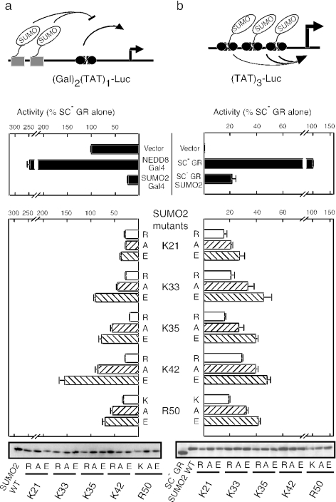
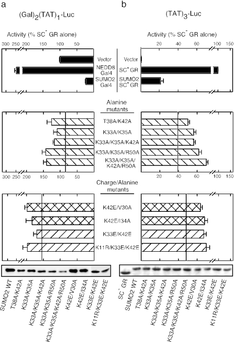
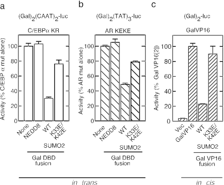
Small ubiquitin-like modifier (SUMO) modification of sequence-specific transcription factors has profound regulatory consequences. By providing an intrinsic inhibitory function, SUMO isoforms can suppress transcriptional activation, particularly at promoters harboring multiple response elements. Through a comprehensive structure-function analysis, we have identified a single critical sector along the second beta sheet and the following alpha helix of SUMO2. This distinct surface is defined by four basic residues (K33, K35, K42, R50) that surround a shallow pocket lined by aliphatic (V30, I34) and polar (T38) residues. Substitutions within this area specifically and dramatically affected the ability of both SUMO2 and SUMO1 to inhibit transcription and revealed that the positively charged nature of the key basic residues is the main feature responsible for their functional role. This highly conserved surface accounts for the inhibitory properties of SUMO on multiple transcription factors and promoter contexts and likely defines the interaction surface for the corepressors that mediate the inhibitory properties of SUMO.


Figures

FIG. 1.
FIG. 1.
Identification of SUMO2 residues involved in repression of GR activity in trans. (a) Effects of single alanine substitutions in SUMO2 on the ability of SUMO2-Gal4 fusions to inhibit ligand-activated SC mutant GR in trans at the pΔ(Gal)2(TAT)1-Luc reporter. Shading of the bars is according to the predicted surface exposure of the corresponding residue. Immunoblot data confirming the expression of the Gal4 fusion proteins are shown below the graph. A schematic representation of GR highlights the two SC/SUMOylation motifs within the N-terminal activation domain (AF-1). The Lys-to-Arg mutations in the SC mutant form are shown below the WT sequence. The DBD and ligand binding domain (LBD) are also indicated. The Δ(Gal)2(TAT)1-Luc reporter is also schematized. Data represent the average ± SEM of at least three independent experiments performed in duplicate or triplicate and are expressed as a percentage of SC mutant GR activity alone (4.9 ± 0.4). (b) Sequence alignment of SUMO and related Ubl proteins. Only residues tested in panel a are shown. Coloring is based on residue type. The locations of secondary-structure elements are shown below the alignment.
FIG. 2.
FIG. 2.
Functionally relevant residues cluster in a distinct region of the surface of SUMO2. Front (a) and back (b) views of ribbon and space-filling representations of the SUMO2 homology model are shown. Coloring of the two leftmost panels is according to the data of Fig. 1 and is normalized to the scale shown below the central panel, ranging from no effect (green) to most deleterious (red). Buried (B) residues predicted to be less than 10% exposed or not tested (NT) are shown in dark and light blue, respectively. In the two rightmost panels, coloring is according to the calculated electrostatic potential from red (negative) to blue (positive) and relative (Rel.) conservation across multiple species, respectively. Residues involved in Ubc9 and SENP2 binding, as well as specific relevant residues, are indicated by the arrows. term, terminus.
FIG. 3.
FIG. 3.
The positive charges of K33, K35, K42, and R50 are critical features for SUMO2-mediated inhibition of GR activity in trans and in cis. (a) Effects of substitutions at select basic residues of SUMO2 on the ability of SUMO2-Gal4 fusions to inhibit SC mutant GR in trans. (b) Effects of the same substitutions on the ability of SUMO2 to inhibit transcription in cis when fused at the N terminus of SC mutant GR. Cells were treated with 10 nM dexamethasone and processed as described in Materials and Methods. Diagrams of the reporter plasmids are shown above the corresponding panels, and expression levels (anti-HA immunoblot) of the fusion proteins are shown below. For both panels a and b, data represent the average ± SEM of at least three independent experiments performed in triplicate and are expressed as a percentage of SC mutant GR activity alone, which was 5.7 ± 0.57 and 190 ± 2 at pΔ(Gal)2(TAT)1-Luc and pΔ(TAT)3-Luc, respectively.
FIG. 4.
FIG. 4.
Compound mutations severely affect SUMO2 inhibition of GR-activated transcription in trans and in cis. (a) Effects of substitutions at multiple positions of SUMO2 on the ability of SUMO2-Gal4 fusions to inhibit SC mutant GR in trans at the pΔ(Gal)2(TAT)1-Luc reporter. (b) Effects of the same substitutions on the ability of SUMO2 to inhibit in cis when fused at the N terminus of SC mutant GR at the pΔTAT3-Luc reporter. Cells were treated with 10 nM dexamethasone and processed as described in Materials and Methods. The reference lines in the middle and bottom parts of each panel indicate the activities of the K42A and K42E mutations, respectively. The expression levels of the corresponding fusions were confirmed by Western blot detection of the HA epitope. In both panels a and b, the data represent the average ± SEM of at least three independent experiments performed in triplicate and are expressed as a percentage of SC mutant GR activity alone, which was 5.7 ± 0.57 and 222 ± 17 at pΔ(Gal)2(TAT)1-Luc and pΔ(TAT)3-Luc, respectively.
FIG. 5.
FIG. 5.
Disruption of the transcriptional regulatory surface of SUMO2 affects inhibition at multiple doses and promoter contexts. (a) Cells were transfected with expression vectors for WT (shaded symbols) or SC mutant (open symbols) GR and the indicated amounts of expression vectors for a Gal4 DBD fusion to either HA-tagged NEDD8 (squares), WT SUMO2 (triangles), or a SUMO2 K33E/K42E mutant (diamonds). The reporter plasmids were pΔ(Gal)2(TAT)1-Luc (left) and pΔ(Gal)2(TAT)3-Luc (right). (b) Cells were transfected with the indicated amounts of p6R-based vectors for the expression of WT GR (shaded circles), SC mutant GR (open squares), or SC mutant GR fused to WT SUMO2 (open triangles) or a SUMO2 K33E/K42E mutant (open diamonds). The reporter plasmids used were pΔTAT1-Luc (left) and pΔTAT3-Luc (right). In both panels a and b, the data represent the average ± SEM of at least three independent experiments performed in triplicate and are expressed as a percentage of SC mutant GR activity alone as follows: pΔ(Gal)2(TAT)1-Luc, 10.8 ± 1.4; pΔ(Gal)2(TAT)3-Luc, 799 ± 100; pΔ(TAT)1-Luc, 3.3 ± 0.2; pΔ(TAT)3-Luc, 471 ± 24.
FIG. 6.
FIG. 6.
Residues implicated in SUMO2-mediated transcriptional repression are functionally conserved in SUMO1. (a) Comparison of the effects of mutations at homologous positions of SUMO1 and SUMO2 on the ability of the corresponding HA-SUMO Gal4 fusions to inhibit SC mutant GR in trans at the pΔ(Gal)2(TAT)1-Luc reporter. (b) Effects of the same mutations on the ability of SUMO1 or -2 to inhibit transcription in cis when fused at the N terminus of SC mutant GR at the pΔTAT3-Luc reporter. In each panel, the data represent the average ± SEM of at least three independent experiments performed in triplicate and are expressed as a percentage of SC mutant GR activity alone either at pΔ(Gal)2(TAT)1-Luc (8.4 ± 1.9) or pΔ(TAT)3-Luc (326 ± 37), respectively.
FIG. 7.
FIG. 7.
The inhibitory surface of SUMO2 influences multiple activators. The ability of the inhibitory surface of SUMO2 to act on multiple activators was tested in trans for SC mutant CEBPα (K159R) from pΔ(Gal)2(CAAT)2-Luc reporter (a) and SC mutant androgen receptor (K385E/K518E) from pΔ(Gal)2(TAT)3-Luc (b)using the indicated Gal4 DBD fusions. (c) In-cis fusions of WT or K33E/K42E SUMO2 to Gal4-VP16 were tested at pΔ(Gal)2-Luc. Cells in panel b were treated with 100 nM dihydrotestosterone. Data represent the average ± SEM of at least three independent experiments performed in triplicate and are expressed as a percentage of the activity of the corresponding activator alone as follows: pΔ(Gal)2(CAAT)2-Luc, 54 ± 2; pΔ(Gal2)2(TAT)3-Luc, 60 ± 5; pΔ(Gal)2-Luc, 300 ± 35. Vec, vector.
FIG. 8.
FIG. 8.
SUMO2 mutants compromised for inhibition are still active for processing and conjugation. (a) The indicated HA-SUMO2 fusions to the Gal4 DBD harboring (+) or lacking (−) the Gly-Gly (GG) motif in the junction between the SUMO2 C terminus (term) and the Gal4 DBD were expressed by transfection into CV-1 cells and tested for processing. Total cell lysates were resolved by SDS-PAGE, and the parental and processed forms were detected by immunoblotting for the HA epitope. (b) In vitro-translated and 35S-labeled SUMO2 proteins lacking the N-terminal SUMOylation site (Δ1-16) alone or in conjunction with the K33E/K42E mutations were tested for interactions with immobilized Ubc9 in a GST pulldown assay as described in Materials and Methods. (c) Pattern of SUMO conjugates in cells expressing HA-tagged and preprocessed (GG stop) WT or mutant (K33E/K42E) SUMO2. Total cell lysates were resolved by SDS-PAGE, and SUMO conjugates were detected by immunoblotting for the HA epitope. Schematic representations of the various SUMO forms used for each experiment are shown. The positions and molecular masses (in kilodaltons) of standards are shown.
FIG. 9.
FIG. 9.
Structural comparison of the repression surface in SUMO1, SUMO2, and SMT3. Front (a) and side (b) views of the transcriptional repression surface in the crystal structures of SUMO2 (left), SUMO1 (center), and SMT3 (right) are shown. The solvent-accessible surface defined by critical functional residues is shown superimposed on the ribbon diagram of each protein. The calculated electrostatic potential is mapped onto each surface, the region occupied by the critical threonine residue (T38 in SUMO2) is shown in yellow, and the position of the hydrophobic hole is outlined with dashed lines. In the side view, the threonine residue is absent to highlight the similar parallel spacing but differing orientation of the critical basic residues in SUMO1 and -2.

Similar articles

Cited by

References

    1. Abdel-Hafiz, H., G. S. Takimoto, L. Tung, and K. B. Horwitz. 2002. The inhibitory function in human progesterone receptor N termini binds SUMO-1 protein to regulate autoinhibition and transrepression. J. Biol. Chem. 277:33950-33956. - PubMed
    1. Alam, S. L., J. Sun, M. Payne, B. D. Welch, B. K. Blake, D. R. Davis, H. H. Meyer, S. D. Emr, and W. I. Sundquist. 2004. Ubiquitin interactions of NZF zinc fingers. EMBO J. 23:1411-1421. - PMC - PubMed
    1. Bailey, D., and P. O'Hare. 2004. Characterization of the localization and proteolytic activity of the SUMO-specific protease, SENP1. J. Biol. Chem. 279:692-703. - PubMed
    1. Bies, J., J. Markus, and L. Wolff. 2002. Covalent attachment of the SUMO-1 protein to the negative regulatory domain of the c-Myb transcription factor modifies its stability and transactivation capacity. J. Biol. Chem. 277:8999-9009. - PubMed
    1. Bohren, K. M., V. Nadkarni, J. H. Song, K. H. Gabbay, and D. Owerbach. 2004. A M55V Polymorphism in a novel SUMO gene (SUMO-4) differentially activates heat shock transcription factors and is associated with susceptibility to type I diabetes mellitus. J. Biol. Chem. 279:27233-27238. - PubMed

Publication types

MeSH terms

LinkOut - more resources