. 2005 May;25(10):4299-310.
doi: 10.1128/MCB.25.10.4299-4310.2005.

The 2 microm plasmid causes cell death in Saccharomyces cerevisiae with a mutation in Ulp1 protease

Affiliations

The 2 microm plasmid causes cell death in Saccharomyces cerevisiae with a mutation in Ulp1 protease

Melanie J Dobson et al. Mol Cell Biol. 2005 May.

Abstract

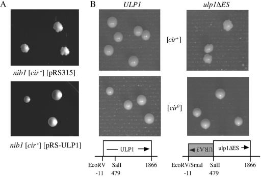
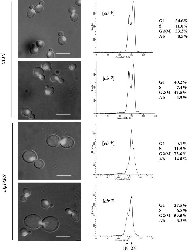
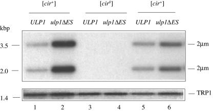
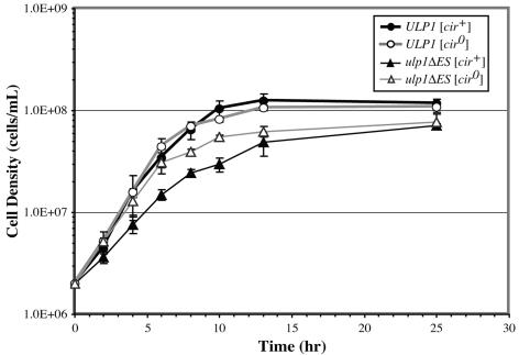
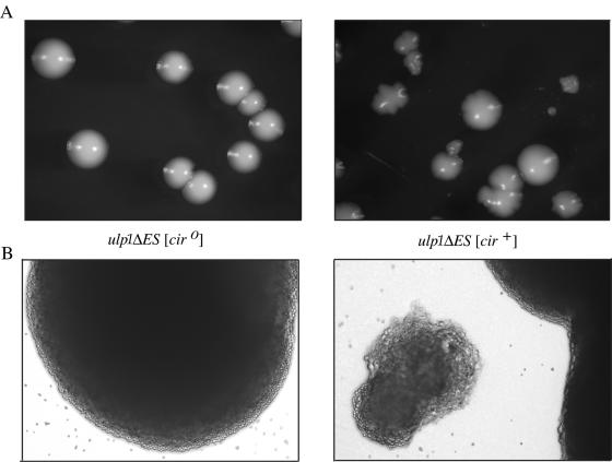
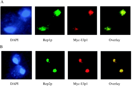
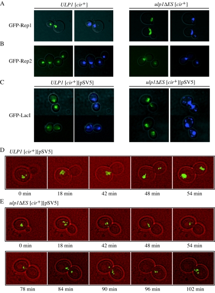
The 2 microm circle plasmid confers no phenotype in wild-type Saccharomyces cerevisiae but in a nib1 mutant, an elevated plasmid copy number is associated with cell death. Complementation was used to identify nib1 as a mutant allele of the ULP1 gene that encodes a protease required for removal of a ubiquitin-like protein, Smt3/SUMO, from protein substrates. The nib1 mutation replaces conserved tryptophan 490 with leucine in the protease domain of Ulp1. Complete deletion of ULP1 is lethal, even in a strain that lacks the 2 microm circle. Partial deletion of ULP1, like the nib1 mutation, results in clonal variations in plasmid copy number. In addition, a subset of these mutant cells produces lineages in which all cells have reduced proliferative capacity, and this phenotype is dependent upon the presence of the 2 microm circle. Segregation of the 2 microm circle requires two plasmid-encoded proteins, Rep1 and Rep2, which were found to colocalize with Ulp1 protein in the nucleus and interact with Smt3 in a two-hybrid assay. These associations and the observation of missegregation of a fluorescently tagged 2 microm circle reporter plasmid in a subset of ulp1 mutant cells suggest that Smt3 modification plays a role in both plasmid copy number control and segregation.


Figures

FIG. 1.
FIG. 1.
Colony morphology of wild-type and nib1 mutant yeasts. (A) Complementation of the nib1 “nibbled” colony morphology by the ULP1 gene. The nib1 [cir+] yeast strain CH569 was transformed with either the vector pRS315 or the plasmid pRS-ULP1, and the transformants were grown on solid SD medium lacking leucine at 30°C for 5 days. (B) Congenic haploid [cir+] and [cir0] yeast strains with either the wild-type ULP1 or mutant ulp1ΔES allele were grown on solid YPD medium at 30°C for 5 days.
FIG. 2.
FIG. 2.
G2/M lag is exacerbated in [cir+] ulp1ΔES yeast. Unsynchronized cultures of congenic ULP1 and ulp1ΔES yeast, [cir+] and [cir0], were grown to early log phase in YPD medium, and the cells were fixed, stained with propidium iodide, visualized by fluorescence microscopy (left), and analyzed by flow cytometry (center), with the percentage of cells at each phase of the cell cycle and the percentage of aberrant cells (Ab), those with DNA content greater than 2N or less than 1N, shown at right. Scale bar, 5 μm.
FIG. 3.
FIG. 3.
Amplification of 2μm circle copy number in ulp1ΔES yeast. Southern blot analysis of EcoRI-restricted genomic DNA isolated from the indicated yeast strains. Phosphorimages of the blots after sequential hybridization with radiolabeled probes recognizing either the genomic TRP1 locus or the 2μm circle (the 2μm circle exists in equimolar amounts of two forms, giving the bands detected with this probe) are shown. TRP1-probed images are longer exposures to allow the weaker signal from this single-copy genomic probe to be visualized.
FIG. 4.
FIG. 4.
The 2μm plasmid impairs growth of ulp1ΔES yeast. Yeast cells were grown to stationary phase in liquid SD medium, diluted into fresh medium, and incubated with shaking at 30°C. Initial and subsequent cell densities at the indicated times after dilution were measured by hemocytometer counts. Results are the averages with standard deviations for four cultures of each strain.
FIG. 5.
FIG. 5.
Variable cell and colony size and morphology in [cir+] ulp1ΔES yeast. (A) [cir0] and [cir+] ulp1ΔES yeast mutants were grown on solid SD medium for 5 days. Colony size and morphology varied only for the [cir+] strain. Several large- and medium-sized nibbled colonies, one large smooth colony, and a small nibbled colony are shown. (B) Bright-field images (magnification, ×100) are of a [cir0] ulp1ΔES colony (left) and [cir+] ulp1ΔES yeast colonies (right), with one small nibbled colony adjacent to a large nibbled colony, from the respective plates above in panel A.
FIG. 6.
FIG. 6.
In situ localization of the Ulp1 and Rep proteins. The Ulp1 protein carrying a carboxy-Myc13 tag was expressed from the native promoter at the ULP1 chromosomal locus. A Myc-specific monoclonal antibody conjugated to rhodamine was used to localize the protein while DNA was stained with DAPI. Native Rep1 (A) and native Rep2 (B) proteins were detected with Rep protein-specific primary polyclonal antibodies and visualized with secondary antibodies conjugated to FITC.
FIG. 7.
FIG. 7.
Localization of Rep proteins and fluorescently tagged 2μm reporter plasmid in [cir+] ULP1 and ulp1ΔES yeast strains. GFP-Rep1 (A), GFP-Rep2 (B), or GFP-LacI (C) fusion proteins were expressed in [cir+] ULP1 and ulp1ΔES yeast strains. The yeasts shown in panel C were also transformed with the 2μm-derived reporter plasmid pSV5, containing lac operator repeats. Merged bright-field-GFP (left) and merged bright-field-DAPI (right) fluorescence images of representative cells are shown. [cir+] ULP1 (D) and [cir+] ulp1ΔES (E) yeast cells expressing GFP-LacI and transformed with pSV5 were analyzed by time-lapse fluorescence microscopy. Bright-field and GFP fluorescence images of a single cell for each strain were taken every 6 min with the small-budded stage taken as the starting time point. Merged bright-field-GFP images are shown for all time points during the period when plasmids moved from mother to daughter cells and from representative earlier and subsequent times.

References

    1. Ahn, Y.-T., X.-L. Wu, S. Biswal, S. Velmurugan, F. C. Volkert, and M. Jayaram. 1997. The 2μm-encoded Rep1 and Rep2 proteins interact with each other and colocalize to the Saccharomyces cerevisiae nucleus. J. Bacteriol. 179:7497-7506. - PMC - PubMed
    1. Bachant, J., A. Alcasabas, Y. Blat, N. Kleckner, and S. J. Elledge. 2002. The SUMO-1 isopeptidase Smt4 is linked to centromeric cohesion through SUMO-1 modification of topisomerase II. Mol. Cell 9:1169-1182. - PubMed
    1. Bartel, P., C. T. Chien, R. Sternglanz, and S. Fields. 1993. Using the two-hybrid system to detect protein-protein interactions., p. 153-179. In D. A. Hartley (ed.), Cellular interactions in development: a practical approach. IRL Press, Oxford, United Kingdom.
    1. Blat, Y., and N. Kleckner. 1999. Cohesins bind to preferential sites along yeast chromosome III, with differential regulation along arms versus the centric region. Cell 98:249-259. - PubMed
    1. Boddy, M. N., K. Howe, L. D. Etkin, E. Solomon, and P. S. Freemont. 1996. Pic-1, a novel ubiquitin-like protein which interacts with the PML component of a multiprotein complex that is disrupted in acute promyelocytic leukaemia. Oncogene 13:971-982. - PubMed

Publication types

MeSH terms

LinkOut - more resources