. 2005 May 23;92(10):1906-14.
doi: 10.1038/sj.bjc.6602593.

Co-expression of RON and MET is a prognostic indicator for patients with transitional-cell carcinoma of the bladder

Affiliations

Co-expression of RON and MET is a prognostic indicator for patients with transitional-cell carcinoma of the bladder

H-L Cheng et al. Br J Cancer. .

Abstract

Recepteur d'Origine Nantais (RON) is a distinct receptor tyrosine kinase in the c-met proto-oncogene family. We examined the mutational and expression patterns of RON in eight human uroepithelial cell lines. Biological effects of RON overexpression on cancer cells were investigated in vitro, and the prognostic significance of RON and/or c-met protein (MET) expression was analysed in a bladder cancer cohort (n=183). There was no evidence of mutation in the kinase domain of RON. Overexpression of RON using an inducible Tet-off system induced increased cell proliferation, motility, and antiapoptosis. Immunohistochemical analysis showed that RON was overexpressed in 60 cases (32.8%) of primary tumours, with 14 (23.3%) showing a high level of expression. Recepteur d'Origine Nantais expression was positively associated with histological grading, larger size, nonpapillary contour, and tumour stage (all P<0.01). In addition, MET was overexpressed in 82 cases (44.8%). Co-expressed RON and MET was significantly associated with decreased overall survival (P=0.005) or metastasis-free survival (P=0.01) in 35 cases (19.1%). Recepteur d'Origine Nantais-associated signalling may play an important role in the progression of human bladder cancer. Evaluation of RON and MET expression status may identify a subset of bladder-cancer patients who require more intensive treatment.


Figures

Figure 1
Figure 1
Immunoprecipitation of MSP in human urine. All nine patients with transitional-cell bladder cancer had detectable MSP in their urine, but MSP was not measurable in the urine of patients with xanthogranulomatous pyelonephritis (case 772). The column of DMEM was taken from medium used for culturing of HepG2 and UB37 cells.
Figure 2
Figure 2
Reverse transcriptase–polymerase chain reaction measurement of RON expression at mRNA level. TCCSUP had the highest level of RON expression in this panel of uroepithelial cell culture, while the other seven cancer cell lines had variable levels of RON expression, with RT4 being the lowest one. Primary culture of uroepithelium was found to have unusually high RON expression. All reactions were performed in triplicate. Data are expressed as relative amounts of RON mRNA levels in relation to PPIA.
Figure 3
Figure 3
Expression of RON and MET receptor proteins in uroepithelial cells. Primary culture of uroepithelial cells and seven cancer cell lines expressed a mature form of p150RON and a precursor p180RON, except that RT cells had very low level of RON expression. All the uroepithelial cells expressed mature p140MET and precursor p170MET, except that TCCSUP cells expressed a relatively lower level of precursor p170MET. These results indicate that co-expression of RON and MET is a universal event in uroepithelial cells.
Figure 4
Figure 4
Growth curve of UB09 cells after transfection of RON. The cells (1 × 10⁴ per well for six wells) were cultured in 10% serum-containing medium with or without doxycycline (1 μg ml−1). The cell number was calculated from 3 to 9 days using a cell counter. Those cells expressing higher RON had a significantly faster growth rate, as indicated by asterisks (P=0.0028, t-test).
Figure 5
Figure 5
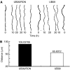
Time-lapse recording of cell migration in UB09 cells after transfection of RON. The cells (5 × 10⁵) were plated on a 1-cm cover slide and grown for 1 day, during which the cells formed a monolayer. Cells were then wounded with a yellow tip. The healing of the wounds was recorded every 20 min for a total of 20 h. (A) Distance of wounding of UB09/RON and UB09 cells, respectively. (B) Quantitative data of migration distance for UB09/RON and UB09 cells, respectively. Overexpression of RON increased the migration distance, symbolic for cell motility, of cancer cells (P=0.04, t-test).
Figure 6
Figure 6
Antiapoptosis by acridine orange staining of the UB09/RON cells after MTX treatment. (A) UB09 cells were electroporated with 40 μg of pLP-TRE2-RON and 40 μg of pTet-Lac-Hyg for 12 h. Cells were first cultured in serum-containing medium for 12 h. Then that medium was replaced with medium containing only MTX (50 μg ml−1), MTX and doxycycline (1 μg ml−1), and, finally, wortmannin (10 nM), MTX, and doxycycline for 60 h each. All the cells were fixed with methanol for 15 min and stained with 1% acridine orange for 10 min. Cells were examined using fluorescent microscopy (left: × 100; right: × 200). Apoptotic cells are indicated by arrows. (B) Levels of apoptotic cells measured using apoptotic body calculation in three different fields were compared in relation to doxycycline or wortmannin treatment (P=0.024, t-test).
Figure 7
Figure 7
Immunohistochemical expression of RON protein in the normal urothelium and bladder cancer cells. (A) Membranous expression of RON receptor was seen in a few non-neoplastic uroepithelial cells (highlighted by arrow). (B) The arrows indicate the membranous staining of RON receptor on cancer cells, representative of a low level of RON expression (original magnification × 300).
Figure 8
Figure 8
Prognostic significance of RON and/or MET expression in bladder cancer patients. Patients who co-expressed RON and MET had a significantly worse metastasis-free survival compared with patients who had single-receptor-positive tumours or no receptor expression (P=0.01).

References

    1. Adam RM, Roth JA, Cheng HL, Rice DC, Khoury J, Bauer SB, Peters CA, Freeman MR (2003) Signaling through PI3K/Akt mediates stretch and PDGF-BB-dependent DNA synthesis in bladder smooth muscle cells. J Urol 169: 2388–2393 - PubMed
    1. Angeloni D, Danilkovitch-Miagkova A, Ivanov SV, Breathnach R, Johnson BE, Leonard EJ, Lerman MI (2000) Gene structure of the human receptor tyrosine kinase RON and mutation analysis in lung cancer samples. Genes Chromosomes Cancer 29: 147–156, doi: 10.1002/1098-2264 (2000)9999:9999〈::AID-GCC1015〉3.0.CO;2-N - PubMed
    1. Chen Q, Seol DW, Carr B, Zarnegar R (1997) Co-expression and regulation of Met and Ron proto-oncogenes in human hepatocellular carcinoma tissues and cell lines. Hepatology 26: 59–66, doi: 10.1002/hep.510260108 - PubMed
    1. Chen YQ, Zhou YQ, Fisher JH, Wang MH (2002) Targeted expression of the receptor tyrosine kinase RON in distal lung epithelial cells results in multiple tumor formation: oncogenic potential of RON in vivo. Oncogene 21: 6382–6386, doi: 10.1038/sj.onc.1205783 - PubMed
    1. Cheng HL, Trink B, Tzai TS, Liu HS, Chan SH, Ho CL, Sidransky D, Chow NH (2002) Overexpression of c-met as a prognostic indicator for transitional cell carcinoma of the urinary bladder. A comparison with p53 nuclear accumulation. J Clin Oncol 20: 1544–1550 - PubMed

Publication types

MeSH terms