. 2005 Jul;77(1):16-26.
doi: 10.1086/431216. Epub 2005 May 3.

Biotin-responsive basal ganglia disease maps to 2q36.3 and is due to mutations in SLC19A3

Affiliations

Biotin-responsive basal ganglia disease maps to 2q36.3 and is due to mutations in SLC19A3

Wen-Qi Zeng et al. Am J Hum Genet. 2005 Jul.

Abstract

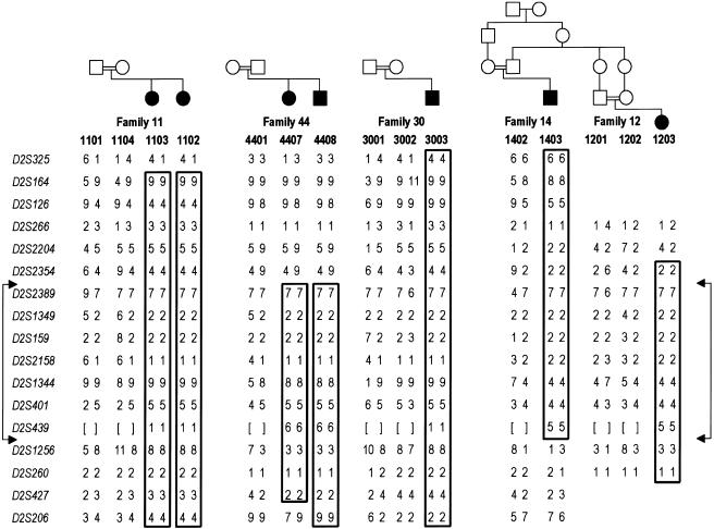
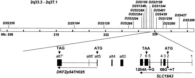
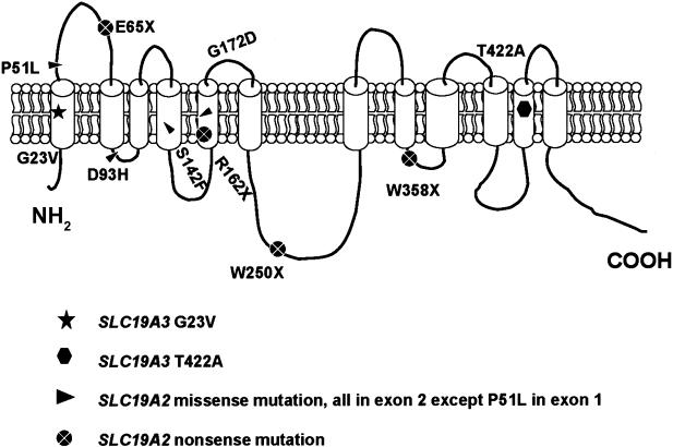
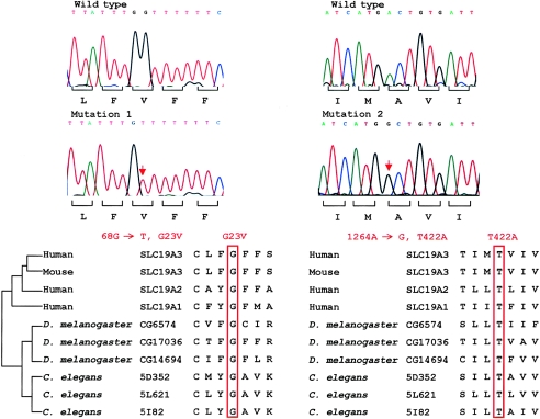
Biotin-responsive basal ganglia disease (BBGD) is a recessive disorder with childhood onset that presents as a subacute encephalopathy, with confusion, dysarthria, and dysphagia, and that progresses to severe cogwheel rigidity, dystonia, quadriparesis, and eventual death, if left untreated. BBGD symptoms disappear within a few days with the administration of high doses of biotin (5-10 mg/kg/d). On brain magnetic resonance imaging examination, patients display central bilateral necrosis in the head of the caudate, with complete or partial involvement of the putamen. All patients diagnosed to date are of Saudi, Syrian, or Yemeni ancestry, and all have consanguineous parents. Using linkage analysis in four families, we mapped the genetic defect near marker D2S2158 in 2q36.3 (LOD=5.9; theta=0.0) to a minimum candidate region (approximately 2 Mb) between D2S2354 and D2S1256, on the basis of complete homozygosity. In this segment, each family displayed one of two different missense mutations that altered the coding sequence of SLC19A3, the gene for a transporter related to the reduced-folate (encoded by SLC19A1) and thiamin (encoded by SLC19A2) transporters.


Figures

Figure  1
Figure 1
Homozygosity mapping and 2q haplotypes in four consanguineous families with BBGD. Blackened symbols indicate patients; unblackened symbols indicate unaffected individuals. Marker alleles, numbered as described in the “Material and Methods” section, are given for seven affected individuals and eight parents within four families with BBGD. Boxes highlight regions of complete homozygosity in affected individuals, indicating disease-chromosome haplotypes. Borders of the minimum candidate interval are denoted by the connected arrows. Genotypes that were not determined are shown by square brackets. Not shown, for confidentiality, are seven unaffected siblings in three of the families who were genotyped and included in the LOD score calculations. None of these samples showed homozygosity for the candidate region. On the basis of the results of mutation analysis, the first three families (11, 20, and 44), all of which involve first-cousin matings, likely share a common ancestor, with the difference in the allele for D2S1344 in family 44 likely being due to a change in the microsatellite marker repeat.
Figure  2
Figure 2
SLC19A3 genomic region, gene structure, and BBGD point mutations. Locations of the microsatellite markers used for the linkage study of the 2q33.3-2q37.1 region and the corresponding gene region for SLC19A3 and DKFZp547H025 are shown. The human SLC19A3 gene is located in 2q36.3, spans 32.91 kb of genomic DNA, and consists of seven exons with a start codon in exon 2 and a TAA stop codon in exon 6. The SLC19A3 cDNA includes a predicted 1,488-bp ORF encoding a 496-aa protein of 55.6 kDa molecular weight. One mutation, 68 G→T, was identified in exon 2, and another mutation, 1264 A→G, was found in exon 5. Sequences alt3, alt4, and alt5 were detected by TBLASTN search (see the BLAST Web site) and were found to be similar to SLC19A3 exons 3, 4, and 5, respectively. The nearby hypothetical protein DKFZp547H025, which is based on a sequenced cDNA, contains three exons (alt5, along with sequences designated “alt6′” and “alt7′,” which are unrelated to SLC19A3), with start and stop codons indicated by arrows. Nine novel SNPs were identified in the 5′ UTR, alt3, alt6′, and alt7′ in SLC19A3 and DKFZp547H025; the SNPs were submitted to dbSNP and were assigned accession numbers 32476166–32476174.
Figure  3
Figure 3
Schematic model of the reduced-folate transporter family. SLC19A3 encodes a predicted protein containing 12 putative transmembrane domains. The relative positions of the mutations identified in SLC19A3 in patients with BBGD and of the missense and nonsense mutations in SLC19A2, reported in patients with Rogers syndrome, are shown (Diaz et al. ; Labay et al. ; Raz et al. ; Scharfe et al. ; Lagarde et al. 2004). The positions of mutations in SLC19A2 were assigned in accordance with hydrophobicity plots that were determined by HMMTOP 2.0 (Tusnady and Simon 1998, 2001). SLC19A2 and SLC19A3 (NCBI accession numbers NP_008927 and NP_079519) were used for analysis.
Figure  4
Figure 4
SLC19A3 point mutations at sites conserved in the reduced-folate transporter family. Sequence chromatograms of the two missense mutations and the corresponding wild-type alleles are shown in the upper panel. These mutations altered glycine (“Mutation 1”) and threonine (“Mutation 2”) residues at codon 23 and codon 422, respectively, that are highly conserved in related transporters in humans, mouse, and lower organisms (lower panel).

References

Web Resources

    1. BLAST, http://www.ncbi.nlm.nih.gov/blast/ (for BLASTX, TBLASTN, and BLASTP)
    1. dbSNP, http://www.ncbi.nlm.nih.gov/SNP/ (for nine novel SNPs in the 5′ UTR, alt3, alt6′, and alt7′ of SLC19A3 and DKFZp547H025 [accession numbers 32476166–32476174])
    1. Fondation Jean Dausset CEPH Web Server, http://www.cephb.fr/ (for CEPH family genotypes used in map construction)
    1. HMMTOP 2.0, http://www.enzim.hu/hmmtop/index.html
    1. Human Genome Database, http://www.gdb.org

References

    1. Aoki Y, Suzuki Y, Sakamoto O, Li X, Takahashi K, Ohtake A, Sakuta R, Ohura T, Miyabayashi S, Narisawa K (1995) Molecular analysis of holocarboxylase synthetase deficiency: a missense mutation and a single base deletion are predominant in Japanese patients. Biochim Biophys Acta 1272:168–174 - PubMed
    1. Balamurugan K, Said HM (2002) Functional role of specific amino acid residues in human thiamine transporter SLC19A2: mutational analysis. Am J Physiol Gastrointest Liver Physiol 283:G37–G43 - PubMed
    1. Baron D, Assaraf YG, Cohen N, Aronheim A (2002) Lack of plasma membrane targeting of a G172D mutant thiamine transporter derived from Rogers syndrome family. Mol Med 8:462–474 - PMC - PubMed
    1. Basel-Vanagaite L, Straussberg R, Ovadia H, Kaplan A, Magal N, Shorer Z, Shalev H, Walsh C, Shohat M (2004) Infantile bilateral striatal necrosis maps to chromosome 19q. Neurology 62:87–90 - PubMed
    1. Buck SH, Burks TF, Brown MR, Yamamura HI (1981) Reduction in basal ganglia and substantia nigra substance P levels in Huntington’s disease. Brain Res 209:464–469 - PubMed

Publication types