The role of magnetic resonance imaging in the assessment of suspected extrinsic tracheobronchial compression due to vascular anomalies
- PMID: 15871985
- PMCID: PMC2083088
- DOI: 10.1136/adc.2004.070250
The role of magnetic resonance imaging in the assessment of suspected extrinsic tracheobronchial compression due to vascular anomalies
Abstract
Aims: To evaluate the role of magnetic resonance imaging (MRI) in the assessment of children with suspected extrinsic tracheobronchial compression due to vascular anomalies.
Methods: Retrospective case note review in a tertiary referral centre. Twenty nine children who underwent dynamic laryngotracheobronchoscopy (DLTB) and were found to have a clinical suspicion of extrinsic tracheobronchial compression were evaluated. All subsequently underwent thoracic MRI within 10 days. The findings on endoscopy were compared to those of MRI, and where performed, echocardiography, aortography, and surgery.
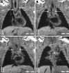
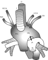
Results: There were 17 males and 12 females (mean age 5 months, range 28 weeks gestation to 60 months). The most common presenting features were stridor and cyanotic episodes. MRI showed abnormalities in 21 patients. There were five vascular rings (three double aortic arches and two right aortic arches) and 11 cases of innominate artery compression. Other vascular anomalies noted included aberrant right subclavian artery and aneurysmal left pulmonary artery. Echocardiography was generally found to be unhelpful in the diagnosis of extra-cardiac vascular abnormalities. Angiography was subsequently conducted in eight children; findings agreed with those shown on MRI. Surgery was performed on all five vascular rings, one innominate artery compression, and one aneurysmal left pulmonary artery. Surgical findings were also compatible with the preoperative MRI.
Conclusions: This study shows the successful use of MRI as the initial imaging modality in endoscopically suspected extrinsic vascular compression of the upper airway. It enables accurate delineation of vascular anomalies and, unlike aortography, is non-invasive and does not require the use of contrast media.
Conflict of interest statement
Competing interests: none
Similar articles
-
Follow-up of surgical correction of aortic arch anomalies causing tracheoesophageal compression: a 38-year single institution experience.J Pediatr Surg. 2009 Jul;44(7):1328-32. doi: 10.1016/j.jpedsurg.2008.11.062. J Pediatr Surg. 2009. PMID: 19573656
-
MR imaging of the pediatric airway.Radiographics. 1995 Mar;15(2):287-98; discussion 298-9. doi: 10.1148/radiographics.15.2.7761634. Radiographics. 1995. PMID: 7761634
-
Vascular anomalies causing tracheoesophageal compression: a 20-year experience in diagnosis and management.Heart Surg Forum. 2003;6(3):149-52. Heart Surg Forum. 2003. PMID: 12821429
-
Vascular tracheobronchial compression syndromes-- experience in surgical treatment and literature review.Thorac Cardiovasc Surg. 2000 Jun;48(3):164-74. doi: 10.1055/s-2000-9633. Thorac Cardiovasc Surg. 2000. PMID: 10903065 Review.
-
Vascular ring abnormalities: a retrospective study of 62 cases.J Pediatr Surg. 2003 Apr;38(4):539-43. doi: 10.1053/jpsu.2003.50117. J Pediatr Surg. 2003. PMID: 12677561 Review.
Cited by
-
CT demonstration of "chicken trachea" resulting from complete cartilaginous rings of the trachea in ring-sling complex.Pediatr Radiol. 2008 Jul;38(7):798-800. doi: 10.1007/s00247-008-0814-0. Epub 2008 Apr 17. Pediatr Radiol. 2008. PMID: 18418589
-
Diagnostic Accuracy of Three-dimensional Turbo Field Echo Magnetic Resonance Imaging Sequence in Pediatric Tracheobronchial Anomalies with Congenital Heart Disease.Sci Rep. 2018 Feb 7;8(1):2529. doi: 10.1038/s41598-018-20892-2. Sci Rep. 2018. PMID: 29416073 Free PMC article.
-
Kommerell's diverticulum in the current era: a comprehensive review.Gen Thorac Cardiovasc Surg. 2015 May;63(5):245-59. doi: 10.1007/s11748-015-0521-3. Epub 2015 Jan 31. Gen Thorac Cardiovasc Surg. 2015. PMID: 25636900 Review.
-
Thoracic aorta aneurysm tracheal compression and anatomical variant of the right subclavian artery: A case report.Radiol Case Rep. 2025 May 14;20(8):3729-3732. doi: 10.1016/j.radcr.2025.04.019. eCollection 2025 Aug. Radiol Case Rep. 2025. PMID: 40486156 Free PMC article.
-
Chronic respiratory disorders due to aberrant innominate artery: a case series and critical review of the literature.Ital J Pediatr. 2023 Jul 22;49(1):92. doi: 10.1186/s13052-023-01473-0. Ital J Pediatr. 2023. PMID: 37480082 Free PMC article. Review.
References
-
- Erwin E A, Gerber M E, Cotton R T. Vascular compression of the airway: indications for and results of surgical management. Int J Pediatr Otorhinolaryngol 199740155–162. - PubMed
-
- Zacharay C H, Myers J L, Eggli K D. Vascular ring due to right aortic arch with mirror‐image branching and left ligamentum arteriosus: complete preoperative diagnosis by magnetic resonance imaging. Pediatr Cardiol 20012271–73. - PubMed
Publication types
MeSH terms
LinkOut - more resources
Full Text Sources
Medical
Molecular Biology Databases


