. 2005 Sep;54(9):1309-17.
doi: 10.1136/gut.2005.065086. Epub 2005 May 4.

CREB/PKA sensitive signalling pathways activate and maintain expression levels of the hepatitis B virus pre-S2/S promoter

Affiliations

CREB/PKA sensitive signalling pathways activate and maintain expression levels of the hepatitis B virus pre-S2/S promoter

F Tacke et al. Gut. 2005 Sep.

Abstract

Background and aims: CREB (cAMP response element binding protein) transcription factors are key regulators of homeostatic functions in the liver, and CRE binding is increased in hepatic inflammation. During chronic hepatitis B virus (HBV) infection, mutations or deletions in the pre-S region are frequently observed. These mutations can affect the pre-S2/S promoter controlling HBV envelope protein expression (hepatitis B surface antigen (HBsAg)) and have been associated with worsened clinical outcome. We aimed to test if CREB activation impacts on HBsAg expression.

Methods: The effect of the CREB inducer protein kinase A (PKA) was tested by coexpression with HBV wild-type vector in vitro. Luciferase reporter gene constructs were cloned to identify novel regulatory regions for the HBV pre-S2/S promoter. Electrophoretic mobility shift assay (EMSA) gelshift and supershift experiments were conducted to confirm DNA transcription factor binding.

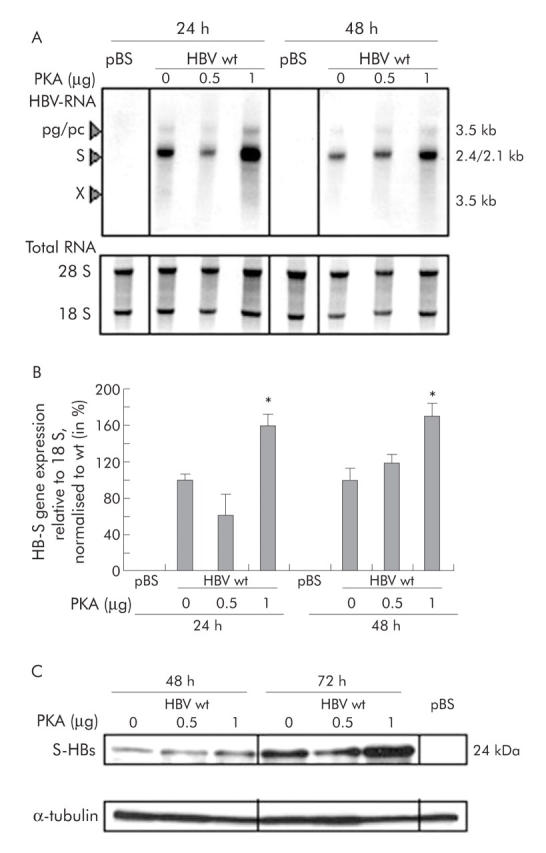
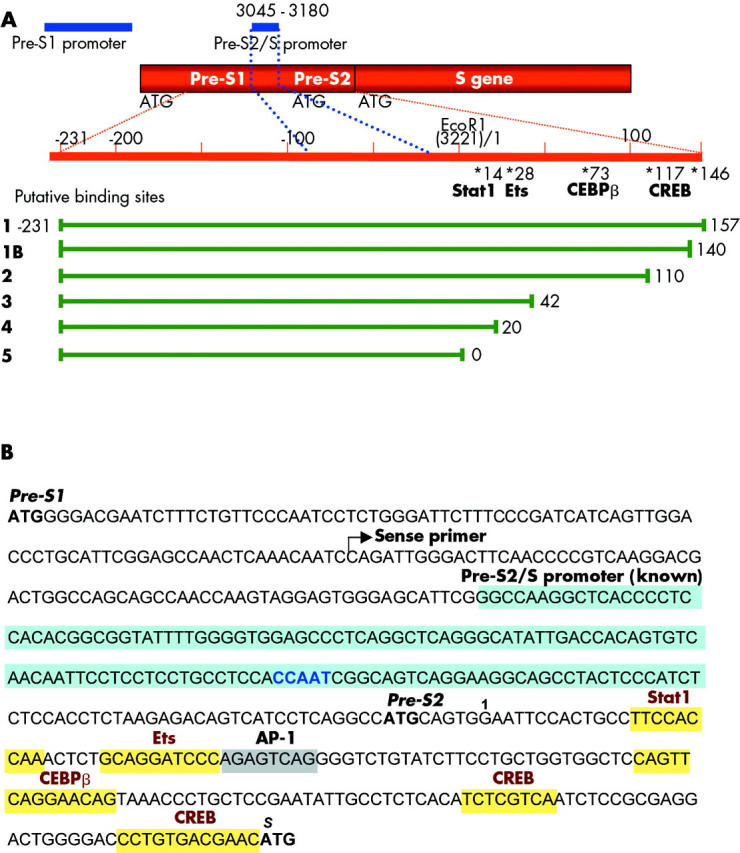
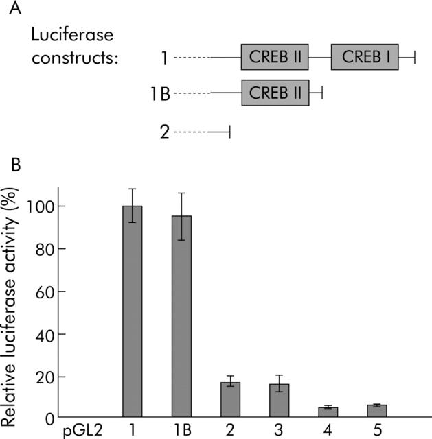
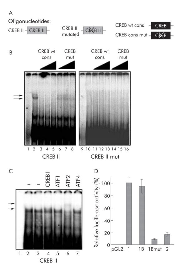
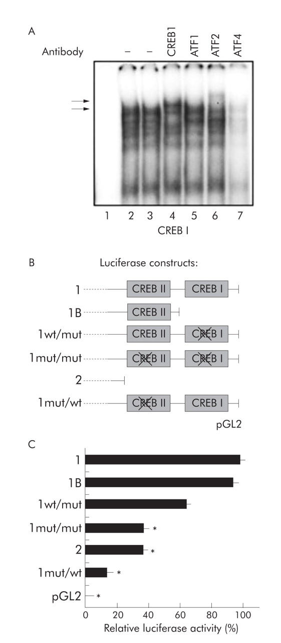
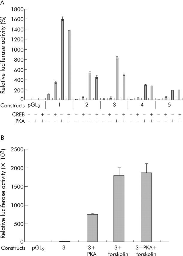
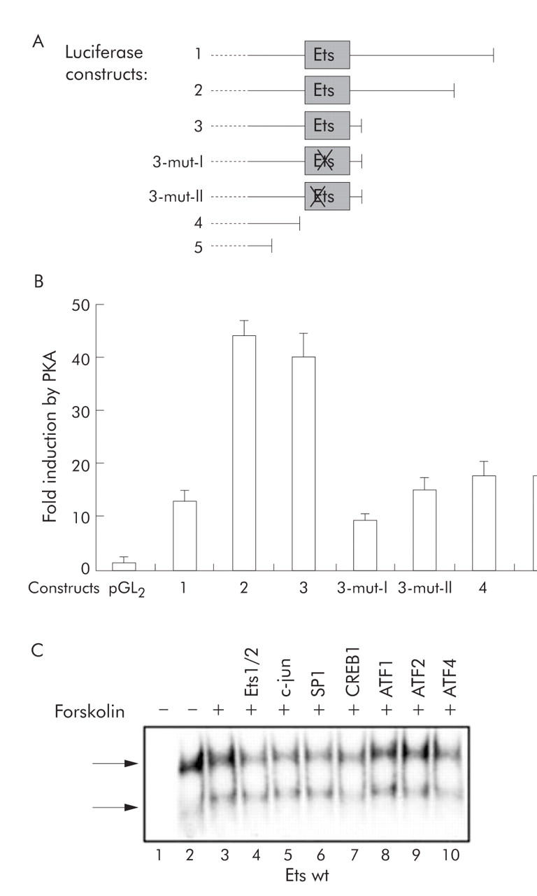
Results: Coexpression of HBV and PKA resulted in HBV-S mRNA induction and enhanced small envelope protein expression. We identified a CREB binding motif in the transcribed part of the pre-S2 region, contributing to basal S promoter activity via binding of activating transcription factor 2 (ATF2). A second CREB motif closely linked to the S-ATG showed a similar binding pattern involving ATF2 and CREB1, without appearing essential for basal promoter activity. Moreover, a sequence in the pre-S2 region is responsible for further transcriptional induction via CREB activators such as PKA and forskolin. EMSA experiments indicate that CREB1 and ATF4 are involved in complex formation conferring PKA dependent promoter activation.

Conclusions: Our data suggest a novel mechanism by which HBV may utilise CREB/PKA signal transduction pathways of hepatocytes to enhance its HBsAg expression during homeostasis and hepatic inflammation.


Figures

Figure 1
Figure 1
Cotransfection of hepatitis B virus (HBV) and the cAMP response element binding protein (CREB) inductor protein kinase A (PKA) leads to increased HBV-S RNA and small hepatitis B surface protein (SHB) expression. (A) Northern Blot analysis. HBV wild-type (wt) plasmid and different concentrations of PKA plasmid were cotransfected. RNA was harvested 24 and 48 hours after transfection, and HBV RNA was detected with a radioactive labelled HBV specific probe. 28S and 18S RNA signals are shown to prove equal loading of RNA. Transfection with pBluescript (pBS) alone served as a negative control. One representative northern blot experiment is shown. pg/pc, pregenomic/precore RNA. (B) Quantification of HBV-S RNA normalised to 18S. Mean (SD) based on three independent experiments. Values are given relative to PKA 0 µg. Significant difference (*p<0.05) compared with PKA 0 µg. (C) Western blot analysis. HBV wild-type (wt) plasmid and different concentrations of PKA plasmid were cotransfected. Total cellular proteins were harvested 48 and 72 hours after transfection, and S-HBs (small HBs protein) was detected with an anti-HBs antibody. Anti-α-tubulin staining of the same blot was performed to prove equal loading. Transfection with pBS served as a negative control. Cotransfection with 1 µg of PKA resulted in increased HBs protein expression.
Figure 2
Figure 2
Organisation of the hepatitis B virus (HBV)-S gene, its promoters, and putative transcription factor binding sites. (A) Schematic diagram of the organisation of the HBV-S gene and its promoters (EcoR1 = 1/3221). The known region of the pre-S2/S promoter (blue) is located within the pre-S1 region from nt 3045–3180. Upstream of the S-ATG, the location of putative transcription factor binding sites for Stat1, Ets, CEBPβ, and cAMP response element binding protein (CREB) are indicated, as revealed by computer based analysis. A putative activator protein 1 (AP-1) site partially overlaps with an Ets motif. Promoter fragments of different lengths (constructs 1–5) were cloned in front of the luciferase reporter gene into the pGL2-plasmid. (B) Sequence of the pre-S1 and pre-S2 region of HBV (genotype A, subtype adw2). ATGs, the start of the applied sense primer (for luciferase constructs) and the known pre-S2/S promoter (blue) with the CCAAT binding motif are highlighted. In addition, upstream binding sites for HNF3 and NF1 plus a downstream binding site for Sp1 have been previously identified to contribute to the known pre-S2/S promoter (not shown). In the pre-S2 region, several transcription factor binding sites were predicted, and are emphasised by yellow boxes.
Figure 3
Figure 3
cAMP response element binding protein (CREB) binding motifs contribute to basal promoter activity. (A) The full length promoter construct 1 (see fig 2A ▶) comprises two putative CREB binding motifs (CREB I/CREB II), as revealed by computer based transcription factor binding search. In addition to the 3′ deletion constructs shown in fig 2A ▶, construct 1B was generated in which the downstream CREB motif (CREB I) is deleted. (B) Luciferase constructs as shown in fig 2A ▶ and fig 3A ▶ were transfected in Huh7 cells and reporter gene expression was quantified (luciferase activity normalised to β-galactosidase activity as an indicator of transfection efficiency). Highest reporter gene expression was seen in construct 1 whereas the 3′ deletion of both putative CREB binding sites as in constructs 2–5 reduced promoter activity below 20%. Deletion of the downstream CREB site alone (construct 1B) did not alter basal promoter activity.
Figure 4
Figure 4
DNA binding analysis for the cAMP response element binding protein (CREB) II motif. (A) Schematic depiction of the oligonucleotides used for the EMSA experiments. CREB II oligo contains the original sequence found in the putative hepatitis B virus (HBV)-S promoter; CREB II mut has a nonsense mutation in the CREB core recognition site. Competition experiments were performed with non-labelled wild-type (wt) or mutated (mut) CREB consensus (cons) oligonucleotides. (B) In EMSA experiments, nuclear extracts from Huh7 cells (except for lanes 1 and 9) were incubated with radioactive labelled double stranded oligonucleotides corresponding to either the wild-type or a mutated sequence of the upstream CREB (CREB II) motif. In lanes 4–6 and 12–14, competition experiments with increasing concentrations of unlabelled (“cold”) consensus wild-type CREB oligo (molar ratios unlabelled:labelled 10:1, 20:1, 40:1), and in lanes 7–8 and 15–16 with cold consensus mutated CREB oligo were performed. Complex formation (arrows) was detected for the wild-type CREB II oligo, but not with the mutated CREB II oligo, that could be specifically reduced by cold CREB consensus wild-type oligonucleotides but not with mutated CREB consensus oligonucleotide. (C) In supershift EMSA experiments, nuclear extracts from Huh7 cells (except for lane 1) were incubated with radioactive labelled double stranded CREB II oligonucleotide and antibodies specific for CREB1 (lane 4), activating transcription factor (ATF) 1 (lane 5), ATF2 (lane 6), and ATF4 (lane 7). Complex formation (lower arrow) could be supershifted with ATF2 antibody (upper arrow). (D) The same mutation from the CREB II mut oligonucleotide, resulting in a lack of specific CREB complex formation, was introduced into the luciferase reporter constructs, and luciferase activity was measured after transfection in Huh7 cells, as described above. Either the mutation (construct 1B mut) or the deletion (as in construct 2) of the upstream CREB II site resulted in a tremendous decrease in luciferase activity compared with constructs 1 and 1B.
Figure 5
Figure 5
Analysis of neighbouring cAMP response element binding protein (CREB) sites for DNA binding specificity and functional redundancy. (A) The neighbouring downstream CREB I site was tested for complex formation in supershift EMSA experiments (as shown in fig 4C ▶ for CREB II). Nuclear extracts from Huh7 cells (except for lane 1) were incubated with radioactive labelled double stranded CREB I oligonucleotide and antibodies specific for CREB1 (lane 4), activating transcription factor (ATF)1 (lane 5), ATF2 (lane 6), and ATF4 (lane 7). Complex formation (lower arrow) could be supershifted with ATF2 and CREB1 antibodies (upper arrow). (B) To determine if the two CREB sites can compensate for each other’s loss of function, additional mutated luciferase constructs were generated as schematically shown bearing nonsense mutations either in one or both CREB motifs. (C) Relative luciferase activity was assessed in transfection experiments. Nonsense mutation of the downstream CREB site (CREB I, construct 1wt/mut) as well as deletion of this motif (construct 1B) resulted only in minor reduction of basal promoter activity whereas deletion (construct 2) or mutation of the upstream CREB site (CREB II, construct 1mut/wt) strongly decreased relative luciferase expression (*p<0.05 compared with construct 1). Introducing mutations of both CREB sites into the full length construct (construct 1mut/mut) had a similar effect.
Figure 6
Figure 6
Identification of a region conferring inducible activity via cAMP response element binding protein (CREB), protein kinase A (PKA), and the exogenous PKA activator forskolin. (A) Deletion constructs 1–5 (see fig 2A ▶) were cotransfected in Huh7 hepatoma cells with 50 ng CREB-plasmid and/or 1 µg PKA expression plasmid, as indicated. Construct 1 conferred the highest basal activity (as expected from figs 3 ▶–5 ▶). Luciferase expression was highly induced by cotransfection of CREB and even further enhanced by PKA in all constructs. However, the highest relative induction was seen in constructs 2 and 3 whereas this inducible activity was remarkably lower in constructs 4 and 5. Constructs 1–3 contain a putative Ets-1 binding motif, which was not present in constructs 4 and 5. (B) Forskolin is a chemical inductor of PKA/CREB signalling pathways. Construct 3 was either cotransfected with PKA and/or cells were stimulated with 10 µM forskolin, as indicated. Stimulation with forskolin resulted in maximal activation of relative luciferase expression.
Figure 7
Figure 7
A putative Ets binding motif mediates inducible activation by protein kinase A (PKA). (A) In order to further characterise the cAMP response element binding protein (CREB)/PKA/forskolin sensitive region, the sequence of the Ets binding motif of construct 3 was mutated (constructs 3-mut-I and 3-mut-II). The constructs used for transfection experiments are shown. (B) Luciferase activity was determined in transfection experiments. Mutations of the Ets motif resulted in reduced inducibility by PKA, similar to the complete deletion of the sequence (constructs 4 and 5). (C) In electrophoretic mobility shift assay (EMSA) experiments, nuclear extracts from either unstimulated Huh7 cells (lane 2) or forskolin stimulated Huh7 cells (lanes 3–10) were incubated with a radioactive labelled double stranded oligonucleotide representing the Ets motif. In order to reduce an unspecific signal (top arrow), nuclear extracts were first preincubated with unlabelled mutated Ets oligonucleotide. Supershift EMSA analysis was performed with antibodies directed against Ets1/2 (lane 4), c-jun (lane 5), SP1 (lane 6), CREB1 (lane 7), activating transcription factor (ATF)1 (lane 8), ATF2 (lane 9), and ATF4 (lane 10). Incubation with anti-CREB1 and anti-ATF4 reduced specific complex formation (bottom arrow, lower signal).

References

    1. Servillo G, Della Fazia MA, Sassone-Corsi P. Coupling cAMP signaling to transcription in the liver: pivotal role of CREB and CREM. Exp Cell Res 2002;275:143–54. - PubMed
    1. Herzig S, Hedrick S, Morantte I, et al. CREB controls hepatic lipid metabolism through nuclear hormone receptor PPAR-gamma. Nature 2003;426:190–3. - PubMed
    1. Herzig S, Long F, Jhala US, et al. CREB regulates hepatic gluconeogenesis through the coactivator PGC-1. Nature 2001;413:179–83. - PubMed
    1. Zhang B, Liu S, Perpetua MD, et al. Cytokines increase CRE binding but decrease CRE-mediated reporter activity in rat hepatocytes by increasing c-Jun. Hepatology 2004;39:1343–52. - PubMed
    1. Mayr B, Montminy M. Transcriptional regulation by the phosphorylation-dependent factor CREB. Nat Rev Mol Cell Biol 2001;2:599–609. - PubMed

Publication types

MeSH terms

Substances