

Respiratory abnormalities resulting from midcervical spinal cord injury and their reversal by serotonin 1A agonists in conscious rats
- PMID: 15872102
- PMCID: PMC6725034
- DOI: 10.1523/JNEUROSCI.5135-04.2005
Respiratory abnormalities resulting from midcervical spinal cord injury and their reversal by serotonin 1A agonists in conscious rats
Abstract
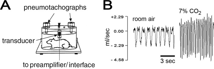
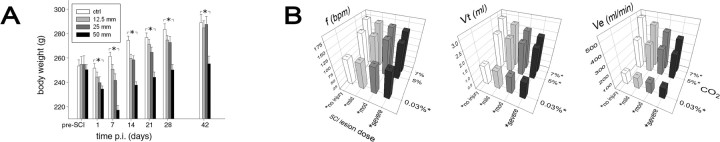
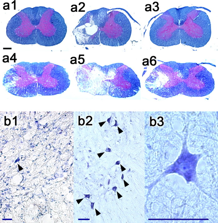
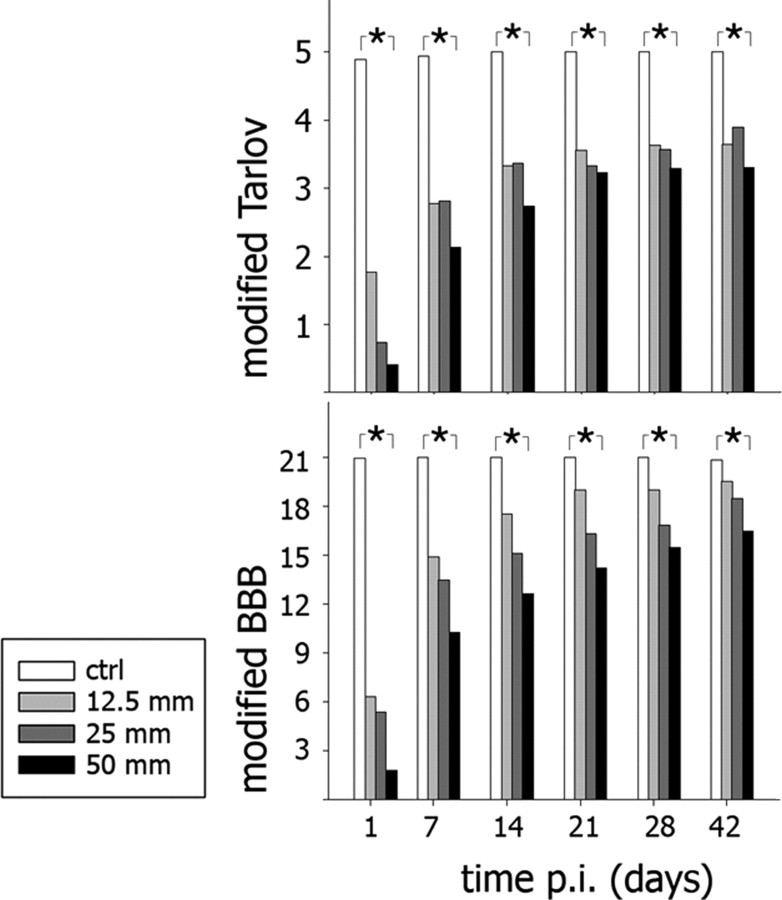
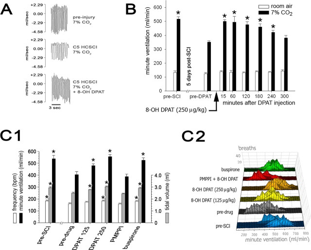
Respiratory dysfunction after cervical spinal cord injury (SCI) has not been examined experimentally using conscious animals, although clinical SCI most frequently occurs in midcervical segments. Here, we report a C5 hemicontusion SCI model in rats with abnormalities that emulate human post-SCI pathophysiology, including spontaneous recovery processes. Post-C5 SCI rats demonstrated deficits in minute ventilation (Ve) responses to a 7% CO2 challenge that correlated significantly with lesion severities (no injury or 12.5, 25, or 50 mm x 10 g weight drop; New York University impactor; p < 0.001) and ipsilateral motor neuron loss (p = 0.016). Importantly, C5 SCI resulted in at least 4 weeks of respiratory abnormalities that ultimately recovered afterward. Because serotonin is involved in respiration-related neuroplasticity, we investigated the impact of activating 5-HT1A receptors on post-C5 SCI respiratory dysfunction. Treatment with the 5-HT1A agonist 8-hydroxy-2-(di-n-propylmino)tetralin (8-OH DPAT) (250 microg/kg, i.p.) restored hypercapnic Ve at 2 and 4 weeks after injury (i.e., approximately 39.2% increase vs post-SCI baseline; p < or = 0.033). Improvements in hypercapnic Ve response after single administration of 8-OH DPAT were dose dependent and lasted for approximately 4 h(p < or = 0.038 and p < or = 0.024, respectively). Treatment with another 5-HT1A receptor agonist, buspirone (1.5 mg/kg, i.p.), replicated the results, whereas pretreatment with a 5-HT1A-specific antagonist, 4-iodo-N-[2-[4(methoxyphenyl)-1-piperazinyl]ethyl]-N-2-pyridinyl-benzamide (3 mg/kg, i.p.) given 20 min before 8-OH DPAT negated the effect of 8-OH DPAT. These results imply a potential clinical use of 5-HT1A agonists for post-SCI respiratory disorders.
Figures
Similar articles
-
Recovery of respiratory activity after C2 hemisection (C2HS): involvement of adenosinergic mechanisms.Respir Physiol Neurobiol. 2009 Nov 30;169(2):102-14. doi: 10.1016/j.resp.2009.07.014. Epub 2009 Aug 3. Respir Physiol Neurobiol. 2009. PMID: 19651244 Free PMC article. Review.
-
Serotonin 1A receptor agonists reverse respiratory abnormalities in spinal cord-injured rats.J Neurosci. 2003 May 15;23(10):4182-9. doi: 10.1523/JNEUROSCI.23-10-04182.2003. J Neurosci. 2003. PMID: 12764106 Free PMC article.
-
Spinal activation of serotonin 1A receptors enhances latent respiratory activity after spinal cord injury.J Spinal Cord Med. 2006;29(2):147-55. doi: 10.1080/10790268.2006.11753868. J Spinal Cord Med. 2006. PMID: 16739558 Free PMC article.
-
Motor deficits and recovery in rats with unilateral spinal cord hemisection mimic the Brown-Sequard syndrome.Brain. 2011 Aug;134(Pt 8):2261-73. doi: 10.1093/brain/awr167. Epub 2011 Jul 13. Brain. 2011. PMID: 21752788
-
Serotonin 1A Receptor Pharmacotherapy and Neuroplasticity in Spinal Cord Injury.Pharmaceuticals (Basel). 2022 Apr 11;15(4):460. doi: 10.3390/ph15040460. Pharmaceuticals (Basel). 2022. PMID: 35455457 Free PMC article. Review.
Cited by
-
Transplantation of Neural Progenitors and V2a Interneurons after Spinal Cord Injury.J Neurotrauma. 2018 Dec 15;35(24):2883-2903. doi: 10.1089/neu.2017.5439. Epub 2018 Aug 10. J Neurotrauma. 2018. PMID: 29873284 Free PMC article.
-
Respiratory neuroplasticity and cervical spinal cord injury: translational perspectives.Trends Neurosci. 2008 Oct;31(10):538-47. doi: 10.1016/j.tins.2008.07.002. Epub 2008 Sep 3. Trends Neurosci. 2008. PMID: 18775573 Free PMC article. Review.
-
Recovery of respiratory activity after C2 hemisection (C2HS): involvement of adenosinergic mechanisms.Respir Physiol Neurobiol. 2009 Nov 30;169(2):102-14. doi: 10.1016/j.resp.2009.07.014. Epub 2009 Aug 3. Respir Physiol Neurobiol. 2009. PMID: 19651244 Free PMC article. Review.
-
Diaphragmatic Activity and Respiratory Function Following C3 or C6 Unilateral Spinal Cord Contusion in Mice.Biology (Basel). 2022 Apr 6;11(4):558. doi: 10.3390/biology11040558. Biology (Basel). 2022. PMID: 35453757 Free PMC article.
-
Time- and dose-related effects of three 5-HT receptor ligands on the genioglossus activity in anesthetized and conscious rats.Sleep Breath. 2007 Dec;11(4):275-84. doi: 10.1007/s11325-007-0107-0. Sleep Breath. 2007. PMID: 17457631
References
-
- Basso DM, Beattie MS, Bresnahan JC (1995) A sensitive and reliable locomotor rating scale for open field testing in rats. J Neurotrauma 12: 1-21. - PubMed
-
- Bjorvatn B, Fagerland S, Eid T, Ursin R (1997) Sleep/waking effects of a selective 5-HT1A receptor agonist given systemically as well as perfused in the dorsal raphe nucleus in rats. Brain Res 770: 81-88. - PubMed
-
- Blier P, Ward NM (2003) Is there a role for 5-HT1A agonists in the treatment of depression? Biol Psychiatry 53: 193-203. - PubMed
-
- Blight AR (1983) Cellular morphology of chronic spinal cord injury in the cat: analysis of myelinated axons by line-sampling. Neuroscience 10: 521-543. - PubMed
-
- Bluechardt MH, Wiens M, Thomas SG, Plyley MJ (1992) Repeated measurements of pulmonary function following spinal cord injury. Paraplegia 30: 768-774. - PubMed
Publication types
MeSH terms
Substances
Grants and funding
LinkOut - more resources
Full Text Sources
Medical
Research Materials
Miscellaneous