. 2005 Sep;25(9):1138-49.
doi: 10.1038/sj.jcbfm.9600121.

Minocycline confers early but transient protection in the immature brain following focal cerebral ischemia-reperfusion

Affiliations

Minocycline confers early but transient protection in the immature brain following focal cerebral ischemia-reperfusion

Christine Fox et al. J Cereb Blood Flow Metab. 2005 Sep.

Erratum in

  • J Cereb Blood Flow Metab. 2006 Mar;26(3):446

Abstract

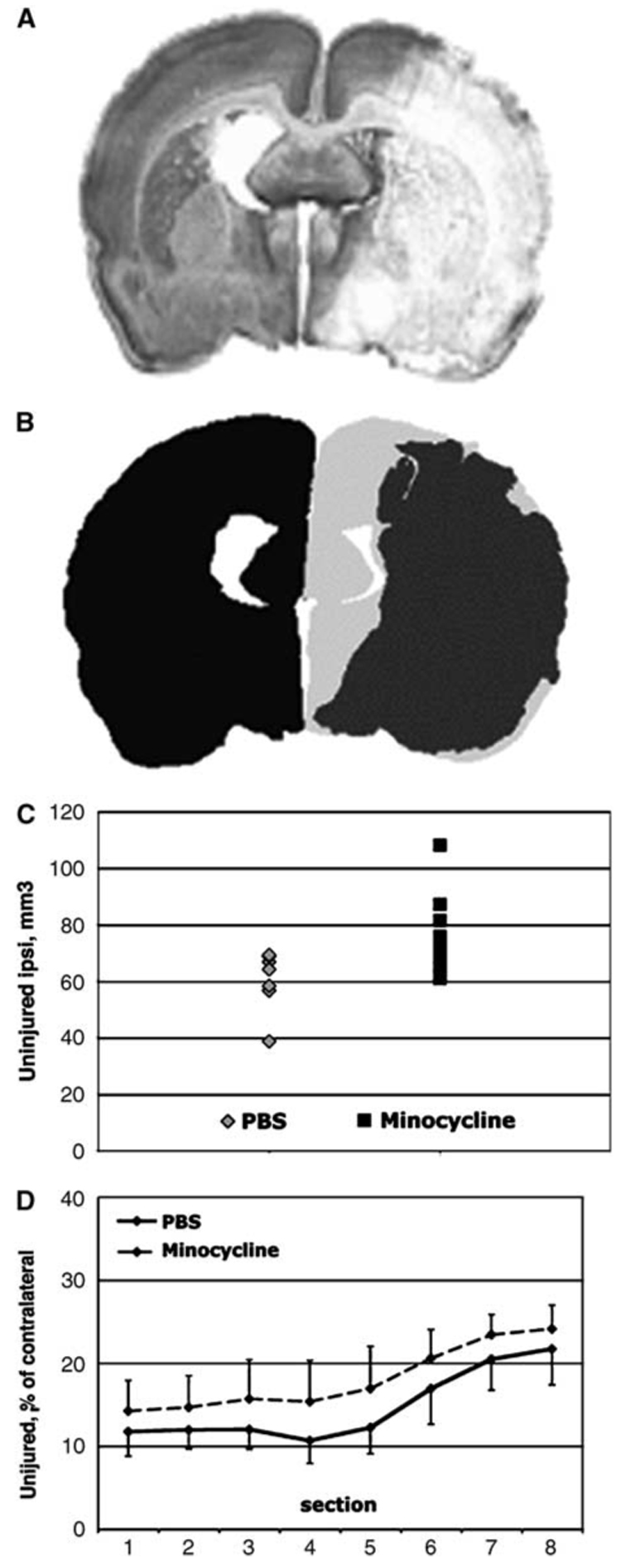
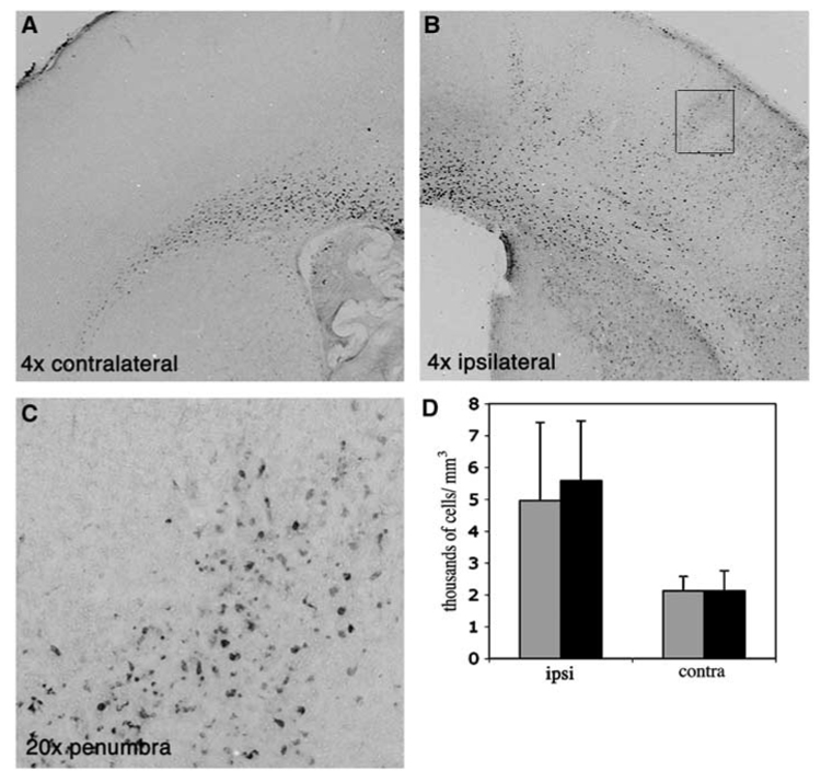
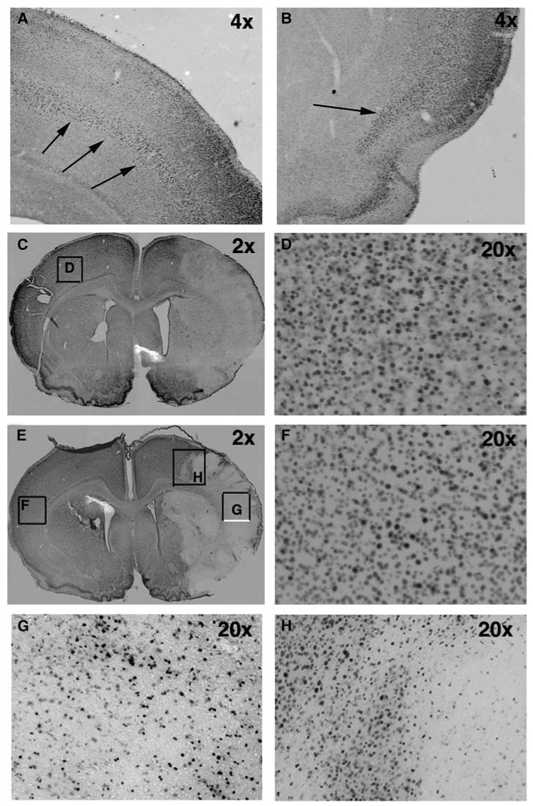
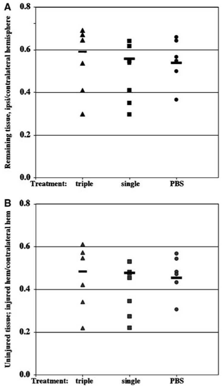
The incidence of neonatal stroke is high and currently there are no strategies to protect the neonatal brain from stroke or reduce the sequelae. Agents capable of modifying inflammatory processes hold promise. We set out to determine whether delayed administration of one such agent, minocycline, protects the immature brain in a model of transient middle cerebral artery (MCA) occlusion in 7-day-old rat pups. Injury volume in minocycline (45 mg/kg/dose, beginning at 2 h after MCA occlusion) and vehicle-treated pups was determined 24 h and 7 days after onset of reperfusion. Accumulation of activated microglia/macrophages, phosphorylation of mitogen-activated protein kinase (MAPK) p38 in the brain, and concentrations of inflammatory mediators in plasma and brain were determined at 24 h. Minocycline significantly reduced the volume of injury at 24 h but not 7 days after transient MCA occlusion. The beneficial effect of minocycline acutely after reperfusion was not associated with changed ED1 phenotype, nor was the pattern of MAPK p38 phosphorylation altered. Minocycline reduced accumulation of IL-1beta and CINC-1 in the systemic circulation but failed to affect the increased levels of IL-1beta, IL-18, MCP-1 or CINC-1 in the injured brain tissue. Therefore, minocycline provides early but transient protection, which is largely independent of microglial activation or activation of the MAPK p38 pathway.


Figures

Figure 1
Figure 1
Minocycline treatment preserves tissue in the ipsilateral hemisphere measured 24 h after MCA occlusion. (A) A representative cresyl violet-stained coronal section demonstrates the morphology of the brain infarct 24 h after MCA occlusion in a control rat pup. Minocycline-treated pups show a similar pattern of injury. (B) Contours are drawn around injured (shown in dark gray) and uninjured (shown in light gray) tissue in the hemisphere ipsilateral to MCA occlusion, as well as around the contralateral hemisphere (shown in black). The size of uninjured tissue is calculated and expressed as a percent of the size of the contralateral hemisphere. (C) The volume of uninjured (unaffected) tissue in the ipsilateral hemisphere is larger (P<0.04) in minocycline-treated than in PBS-treated pups. Each dot represents the size of uninjured tissue for an individual pup. (D) Minocycline treatment consistently preserves the tissue in all eight studied sections in the anterior to posterior direction.
Figure 2
Figure 2
Accumulation of ED1 immunoreactive cells in the injured hemisphere is not affected by minocycline treatment 24 h after MCA occlusion. Representative examples of ED1 immunostained sections contralateral (A) and ipsilateral (B, C) to MCA occlusion. (C) shows a higher magnification picture (20 ×) of the frame shown in (B). While in the uninjured hemisphere the presence of ED1-positive cells is limited to corpus collosum (A), rapid acquisition of ED1 phenotype is evident in the lesioned tissue (B, C). (D) Density of ED1 immunoreactive cells in minocycline and PBS-treated pups 24 h after reperfusion. Black—PBS-treated, Gray—minocycline-treated pups.
Figure 3
Figure 3
The pattern of the expression of phosphorylated form of MAP kinase p38 is not affected by minocycline treatment at 8 h after reperfusion. (A, B) Expression of p-p38 in a naïve brain of P7 rat. Arrows demonstrate the laminar pattern of p38 phosphorylation. (C) The pattern of p38 phosphorylation following PBS treatment (2 ×). A unilateral decrease of p38 phosphorylation is observed in injured but not in unaffected brain tissue. (D) A higher magnification picture obtained from the box in panel C. (E) The pattern of p38 phosphorylation following minocycline treatment (2 ×). (F) A higher magnification picture obtained from the box in panel E. (G) A higher magnification picture (20 ×, box in panel E) shows that p38 phosphorylation is seen only in a subpopulation of cells in the ischemic core. (H) A higher magnification picture (20 ×, box in panel E) of p38 phosphorylation. A heterogeneous pattern of p38 phosphorylation is observed in the ischemic penumbra 8 h post-reperfusion.
Figure 4
Figure 4
Minocycline treatment does not change the volume of the remaining and uninjured tissue of lesioned hemisphere 7 days after transient MCA occlusion. Volumes of the contralateral hemisphere, remaining ipsilateral and morphologically preserved ipsilateral hemisphere were determined in eight consecutive 15-µm-thick paraffin-embedded cresyl violet-stained coronal sections. Sections were obtained at 400-µm intervals. Volumes of the remaining ipsilateral (A) and morphologically normal/unaffected (B) tissues were normalized to the volume of contralateral hemisphere in serial sections. Pups treated with a single or triple minocycline injections show no difference in the volume of the remaining (A) or preserved (B) tissue in the hemisphere ipsilateral to the occlusion.

References

    1. Arvin KL, Han BH, Du Y, Lin SZ, Paul SM, Holtzman DM. Minocycline markedly protects the neonatal brain against hypoxic–ischemic injury. Ann Neurol. 2002;52:54–61. - PubMed
    1. Barone FC, Feuerstein GZ. Inflammatory mediators and stroke: new opportunities for novel therapeutics. J Cereb Blood Flow Metab. 1999;19:819–834. - PubMed
    1. Barone FC, Irving EA, Ray AM, Lee JC, Kassis S, Kumar S, Badger AM, White RF, McVey MJ, Legos JJ, Erhardt JA, Nelson AH, Ohlstein EH, Hunter AJ, Ward K, Smith BR, Adams JL, Parsons AA. SB 239063, a second-generation p38 mitogen-activated protein kinase inhibitor, reduces brain injury and neurological deficits in cerebral focal ischemia. J Pharmacol Exp Ther. 2001;296:312–321. - PubMed
    1. Benjelloun N, Renolleau S, Represa A, Ben-Ari Y, Charriaut-Marlangue C. Inflammatory responses in the cerebral cortex after ischemia in the P7 neonatal rat. Stroke. 1999;30:1916–1923. discussion 1923–1914. - PubMed
    1. Bhat NR, Zhang P, Lee JC, Hogan EL. Extracellular signal-regulated kinase and p38 subgroups of mitogen-activated protein kinases regulate inducible nitric oxide synthase and tumor necrosis factor-alpha gene expression in endotoxin-stimulated primary glial cultures. J Neurosci. 1998;18:1633–1641. - PMC - PubMed

Publication types

MeSH terms