

PKC translocation and ERK1/2 activation in compensated right ventricular hypertrophy secondary to chronic emphysema
- PMID: 15876346
- PMCID: PMC1142330
- DOI: 10.1186/1472-6793-5-6
PKC translocation and ERK1/2 activation in compensated right ventricular hypertrophy secondary to chronic emphysema
Abstract
Background: Right ventricular hypertrophy (RVH) is an important complication of chronic lung disease. However, the signal transduction pathways involved as well as the physiological changes to the right ventricle have not been investigated. Emphysema was produced in male, Syrian Golden hamsters by intra-tracheal instillation of 250 IU/kg elastase (Emp, n = 17). Saline treated animals served as controls (Con, n = 15).
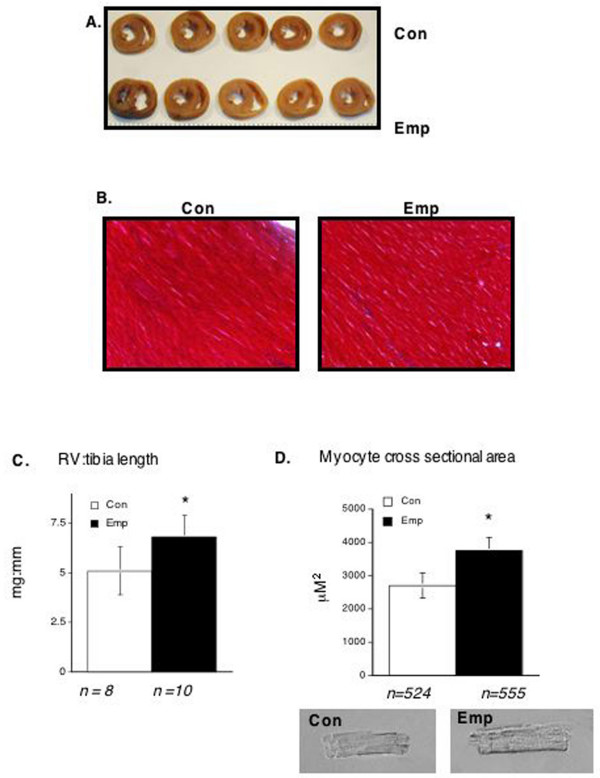
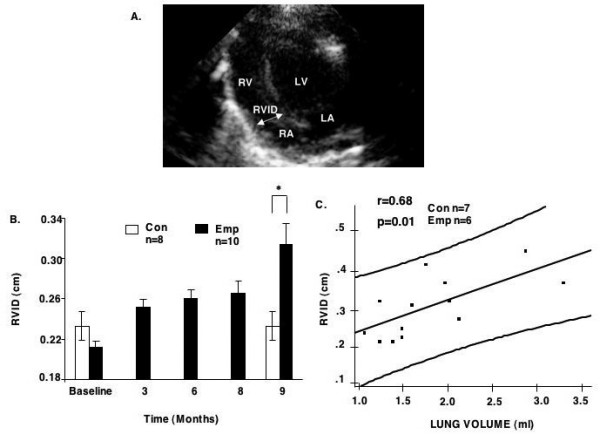
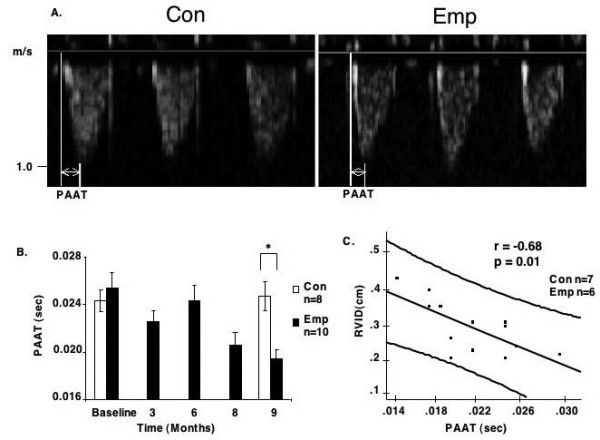
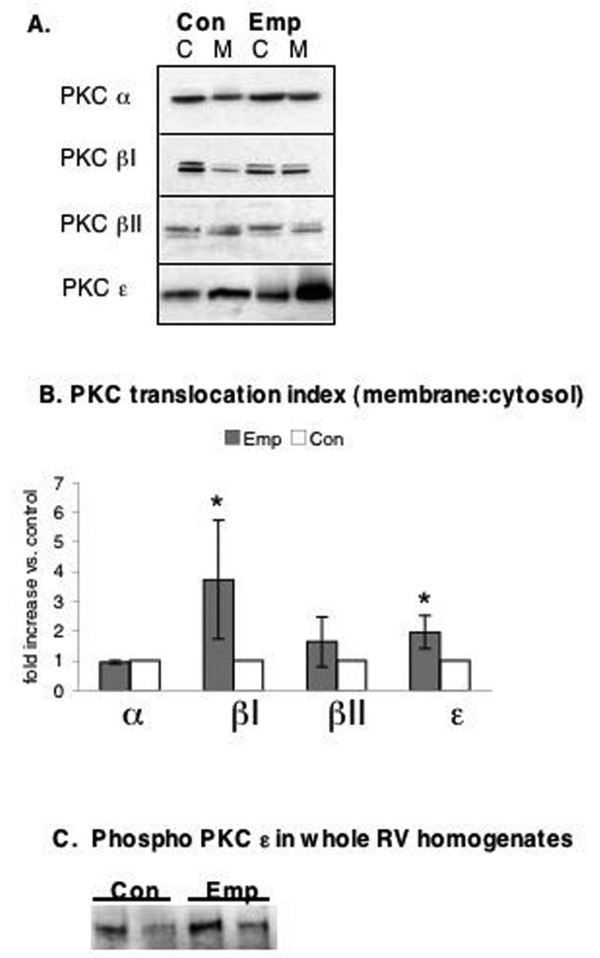
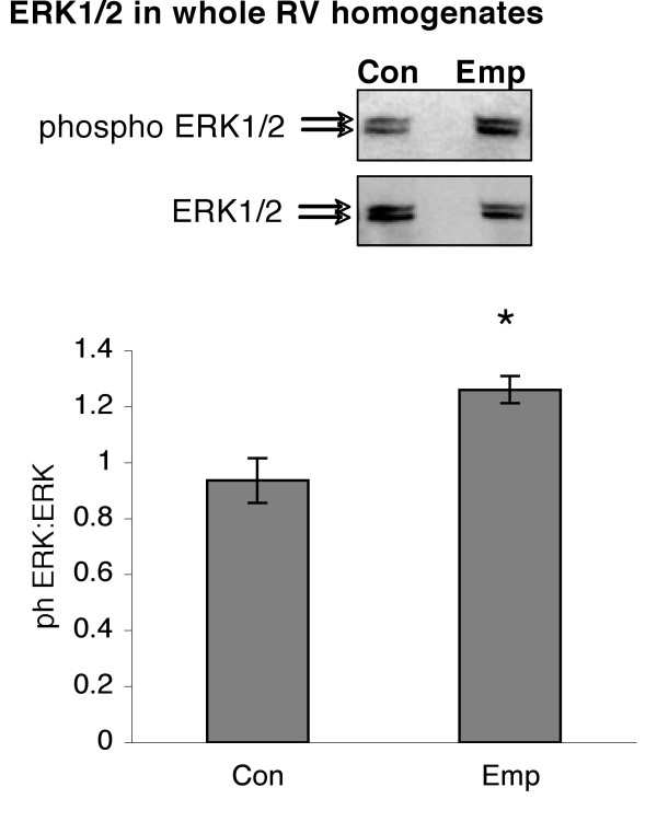
Results: Nine months later, Emp hamsters had 75% greater lung volume, and evidence of RVH at the gross and myocyte level (RV:tibia length Emp 6.84 +/- 1.18 vs. Con 5.14 +/- 1.11 mg/mm; myocyte cross sectional area Emp 3737 vs. Con 2695 microm2), but not left ventricular hypertrophy. Serial echocardiographic analysis from baseline to nine months after induction of emphysema revealed increasing right ventricular internal dimension and decreased pulmonary artery acceleration time only in Emp hamsters. There was an increase in translocation of PKC betaI and PKC epsilon from cytosolic to membranous cell fractions in RV of Emp hamsters. Phosphorylation of PKC epsilon was unchanged. Translocation of PKC alpha and betaII were unchanged. Emp animals had a 22% increase in phospho-ERK 1/2, but no change in levels of total ERK 1/2 compared to Con.
Conclusion: These data suggest that PKC betaI, epsilon and ERK 1/2 may play a role in mediating compensated RVH secondary to emphysema and may have clinical relevance in the pathogenesis of RVH.
Figures
Similar articles
-
Protein kinase C isozyme expression in right ventricular hypertrophy induced by pulmonary hypertension in chronically hypoxic rats.Mol Med Rep. 2017 Oct;16(4):3833-3840. doi: 10.3892/mmr.2017.7098. Epub 2017 Jul 27. Mol Med Rep. 2017. PMID: 28765942 Free PMC article.
-
Bosentan attenuates right ventricular hypertrophy and fibrosis in normobaric hypoxia model of pulmonary hypertension.J Heart Lung Transplant. 2011 Jul;30(7):827-33. doi: 10.1016/j.healun.2011.03.010. Epub 2011 May 8. J Heart Lung Transplant. 2011. PMID: 21550822 Free PMC article.
-
Right ventricular hypertrophy and apoptosis after pulmonary artery banding: regulation of PKC isozymes.Cardiovasc Res. 2003 Sep 1;59(3):658-67. doi: 10.1016/s0008-6363(03)00470-x. Cardiovasc Res. 2003. PMID: 14499867
-
Protein kinase C (PKC)-delta/-epsilon mediate the PKC/Akt-dependent phosphorylation of extracellular signal-regulated kinases 1 and 2 in MCF-7 cells stimulated by bradykinin.J Endocrinol. 2006 Jan;188(1):79-89. doi: 10.1677/joe.1.06433. J Endocrinol. 2006. PMID: 16394177
-
Protein kinase C-alpha-induced hypertrophy of neonatal rat ventricular myocytes.Am J Physiol Heart Circ Physiol. 2004 Dec;287(6):H2777-89. doi: 10.1152/ajpheart.00171.2004. Epub 2004 Jul 22. Am J Physiol Heart Circ Physiol. 2004. PMID: 15271671
Cited by
-
Effects of the association of diabetes and pulmonary emphysema on cardiac structure and function in rats.Int J Exp Pathol. 2015 Oct;96(5):350-7. doi: 10.1111/iep.12146. Epub 2015 Oct 29. Int J Exp Pathol. 2015. PMID: 26515722 Free PMC article.
-
Cytosolic, but not mitochondrial, oxidative stress is a likely contributor to cardiac hypertrophy resulting from cardiac specific GLUT4 deletion in mice.FEBS J. 2012 Feb;279(4):599-611. doi: 10.1111/j.1742-4658.2011.08450.x. Epub 2012 Jan 9. FEBS J. 2012. PMID: 22221582 Free PMC article.
-
Mammalian target of rapamycin is a critical regulator of cardiac hypertrophy in spontaneously hypertensive rats.Hypertension. 2009 Dec;54(6):1321-7. doi: 10.1161/HYPERTENSIONAHA.109.138818. Epub 2009 Nov 2. Hypertension. 2009. PMID: 19884565 Free PMC article.
-
A low-carbohydrate/high-fat diet reduces blood pressure in spontaneously hypertensive rats without deleterious changes in insulin resistance.Am J Physiol Heart Circ Physiol. 2013 Jun 15;304(12):H1733-42. doi: 10.1152/ajpheart.00631.2012. Epub 2013 Apr 19. Am J Physiol Heart Circ Physiol. 2013. PMID: 23604708 Free PMC article.
References
-
- Lucas JW SJSBV. National Center for Health Statistics, Vital Health Statistics. Vol. 10. Hyattsville, MD , National Center for Health Statistics; 2004. Summary health statistics for U.S. Adults: National Health Interview Survey, 2001. - PubMed
-
- Vizza CD, Lynch JP, Ochoa LL, Richardson G, Trulock EP. Right and left ventricular dysfunction in patients with severe pulmonary disease. Chest. 1998;113:576–583. - PubMed
-
- Hayes JA, Christensen TG, Snider GL. The hamster as a model of chronic bronchitis and emphysema in man. Lab Anim Sci. 1977;27:762–770. - PubMed
Publication types
MeSH terms
Substances
Grants and funding
LinkOut - more resources
Full Text Sources
Medical
Miscellaneous
