. 2005 May 6:6:18.
doi: 10.1186/1471-2350-6-18.

Molecular breakpoint cloning and gene expression studies of a novel translocation t(4;15)(q27;q11.2) associated with Prader-Willi syndrome

Affiliations

Molecular breakpoint cloning and gene expression studies of a novel translocation t(4;15)(q27;q11.2) associated with Prader-Willi syndrome

Birgitt Schüle et al. BMC Med Genet. .

Abstract

Background: Prader-Willi syndrome (MIM #176270; PWS) is caused by lack of the paternally-derived copies, or their expression, of multiple genes in a 4 Mb region on chromosome 15q11.2. Known mechanisms include large deletions, maternal uniparental disomy or mutations involving the imprinting center. De novo balanced reciprocal translocations in 5 reported individuals had breakpoints clustering in SNRPN intron 2 or exon 20/intron 20. To further dissect the PWS phenotype and define the minimal critical region for PWS features, we have studied a 22 year old male with a milder PWS phenotype and a de novo translocation t(4;15)(q27;q11.2).

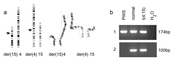
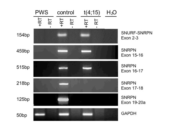
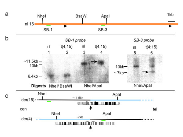
Methods: We used metaphase FISH to narrow the breakpoint region and molecular analyses to map the breakpoints on both chromosomes at the nucleotide level. The expression of genes on chromosome 15 on both sides of the breakpoint was determined by RT-PCR analyses.

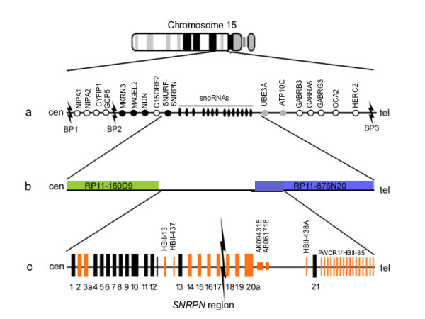
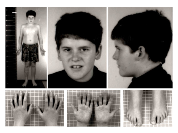
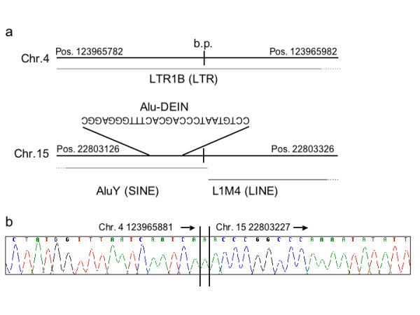
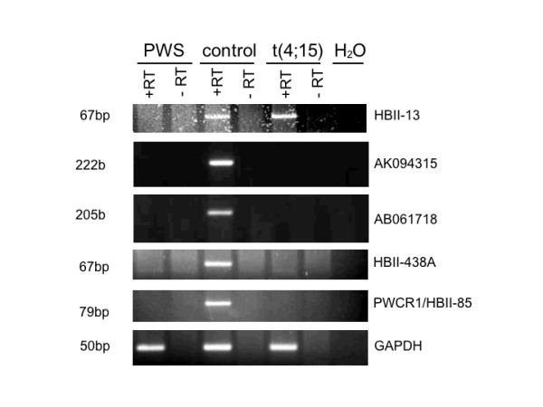
Results: Pertinent clinical features include neonatal hypotonia with feeding difficulties, hypogonadism, short stature, late-onset obesity, learning difficulties, abnormal social behavior and marked tolerance to pain, as well as sticky saliva and narcolepsy. Relative macrocephaly and facial features are not typical for PWS. The translocation breakpoints were identified within SNRPN intron 17 and intron 10 of a spliced non-coding transcript in band 4q27. LINE and SINE sequences at the exchange points may have contributed to the translocation event. By RT-PCR of lymphoblasts and fibroblasts, we find that upstream SNURF/SNRPN exons and snoRNAs HBII-437 and HBII-13 are expressed, but the downstream snoRNAs PWCR1/HBII-85 and HBII-438A/B snoRNAs are not.

Conclusion: As part of the PWCR1/HBII-85 snoRNA cluster is highly conserved between human and mice, while no copy of HBII-438 has been found in mouse, we conclude that PWCR1/HBII-85 snoRNAs is likely to play a major role in the PWS- phenotype.


Figures

Figure 1
Figure 1
Mapping the t(4;15) breakpoint and expression patterns of SNRPN exons and intronic genes . a . Schematic map of human chromosome region 15q11-q13. Black and gray circles represent imprinted genes, expressed from the paternal or maternal allele, respectively. White circles designate bi-allelically expressed genes. BP1, 2, and 3 indicate the locations of the deletion breakpoint hotspots [43]. b . FISH results placed BAC RP11-160D9 highlighted in green (nucleotide position 22577151-22735621) proximal to the translocation breakpoint and RP11-876N20 highlighted in blue (position 22857334-23036552) distal to the breakpoint. Intron 17, comprising nucleotides 22795282 to 22811656, thus is located ~ 63.4 kb downstream of RP11-160D9 and ~ 42 kb upstream of RP11-876N20. c . On representation of the SNRPN region (not drawn to scale) boxes represent exons and ESTs, lines represent snoRNA copies. Orange boxes and lines indicate exons, ESTs or snoRNAs tested for expression either by RT-PCR or quantitative RT-PCR. Black flash indicates the breakpoint in intron 17 of the SNRPN locus.
Figure 2
Figure 2
t(4;15) carrier at 15 years of age . Note absence of typical PWS facial features and presence of mild truncal obesity.
Figure 3
Figure 3
a. High resolution G-banded ideograms and prometaphase chromosomes of the translocation derivatives and their normal homologs . An apparently balanced translocation t(4;15)(q27;q11) was identified with arrows indicating band location of breakpoints. b. DNA methylation analysis of CpG island of SNRPN promoter and exon 1. 1. The 174 bp PCR product is derived from the methylated maternal chromosome. 2. The 100 bp product is derived from the paternal chromosome. PWS: PWS control, Normal: normal control, and t(4;15) carrier; H2O: no template control. The t(4;15) carrier shows the normal bi-parental methylation pattern.
Figure 4
Figure 4
SNRPN expression analysis by RT-PCR of RNA from LCLs . On the left, the sizes of the PCR products are shown, and on the right, the location of the primers in SNRPN exons is listed. +RT: with reverse transcriptase; -RT: without reverse transcriptase; H2O: no template control. All SNRPN +RT products tested were absent in the PWS control, and present in the normal control. The t(4;15) cells were positive for SNURF/ SNRPN exons 2–3, 15–16 and 16–17 and negative for exons 18 through 20a. GAPDH primers were used as control for the integrity of the cDNA.
Figure 5
Figure 5
Southern blot analysis identifies breakpoint in SNRPN intron 17 . a . Restriction map of the intron 17 region of the SNRPN gene on the normal chromosome 15. Black arrowheads indicate the boundaries of intron 17. The positions of the two hybridization probes (SB-1 and SB-3) are indicated by green lines. b . Lanes 1 and 2 contain double digests with NheI and BsaWI to release a fragment of 6.4 kb, lanes 3 and 4 contain double digests with NheI and ApaI to release a fragment of 10 kb. The membrane was probed with probe SB-1. The arrow indicates an additional band above the 10 kb fragment ~11.5 kb in length. The two bands are not well resolved on the rendition of this blot. This novel fragment is represented in c, upper panel. Lanes 5 and 6 contain double digests with NheI and ApaI to release a 10 kb fragment. The membrane is probed with SB-3. The arrow indicates an additional band of ~ 7 kb. This novel fragment is represented schematically in c, lower panel. c . Schematic representation of the junction fragments identified on the Southern blot in b. The upper panel represents the der(15) and the lower panel represents the der(4). Chromosome 15 material is indicated as a black line and material from chromosome 4 as a blue line. Location of restriction sites and of hybridization probes (green lines) are indicated.
Figure 6
Figure 6
Repeat sequences surrounding the breakpoint . a . One hundred nucleotides on either side of the breakpoints on chromosome 4 and 15 contain repetitive sequences (grey lines). The Alu-DEIN sequence is located 13–39 bp upstream of the breakpoint on chromosome 15. b . Sequence across the breakpoint on the der(4) chromosome reveals an additional A inserted at the breakpoint. Arrows indicate the direction centromere to telomere.
Figure 7
Figure 7
Expression analysis in LCLs of snoRNAs and two ESTs in intron 20 . RT-PCR analysis of the C/D box snoRNAs reveals expression of HBII-13, but not of HBII-438A/B, PWCR1/HBII-85 and the two ESTs in intron 20 in the t(4;15) translocation carrier. +RT: with reverse transcriptase; -RT: without reverse transcriptase; H2O: no template control.

References

    1. Holm VA, Cassidy SB, Butler MG, Hanchett JM, Greenswag LR, Whitman BY, Greenberg F. Prader-Willi syndrome: consensus diagnostic criteria. Pediatrics. 1993;91:398–402. - PMC - PubMed
    1. Cassidy SB. Prader-Willi syndrome. 2001. pp. 301–322.
    1. Ledbetter DH, Riccardi VM, Airhart SD, Strobel RJ, Keenan BS, Crawford JD. Deletions of chromosome 15 as a cause of the Prader-Willi syndrome. N Engl J Med. 1981;304:325–329. - PubMed
    1. Buiting K, Gross S, Lich C, Gillessen-Kaesbach G, el-Maarri O, Horsthemke B. Epimutations in Prader-Willi and Angelman syndromes: a molecular study of 136 patients with an imprinting defect. Am J Hum Genet. 2003;72:571–577. doi: 10.1086/367926. - DOI - PMC - PubMed
    1. Mascari MJ, Gottlieb W, Rogan PK, Butler MG, Waller DA, Armour JA, Jeffreys AJ, Ladda RL, Nicholls RD. The frequency of uniparental disomy in Prader-Willi syndrome. Implications for molecular diagnosis. N Engl J Med. 1992;326:1599–1607. - PMC - PubMed

Publication types

MeSH terms