Comparative Study
. 2005 May 17;102(20):7163-8.
doi: 10.1073/pnas.0502164102. Epub 2005 May 6.

Structural and functional similarities between the capsid proteins of bacteriophages T4 and HK97 point to a common ancestry

Affiliations
Comparative Study

Structural and functional similarities between the capsid proteins of bacteriophages T4 and HK97 point to a common ancestry

Andrei Fokine et al. Proc Natl Acad Sci U S A. .

Abstract

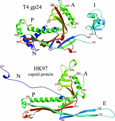
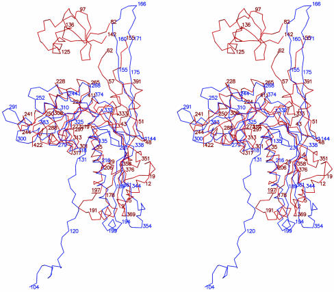
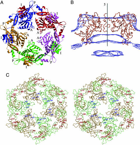
Gene product (gp) 24 of bacteriophage T4 forms the pentameric vertices of the capsid. Using x-ray crystallography, we found the principal domain of gp24 to have a polypeptide fold similar to that of the HK97 phage capsid protein plus an additional insertion domain. Fitting gp24 monomers into a cryo-EM density map of the mature T4 capsid suggests that the insertion domain interacts with a neighboring subunit, effecting a stabilization analogous to the covalent crosslinking in the HK97 capsid. Sequence alignment and genetic data show that the folds of gp24 and the hexamer-forming capsid protein, gp23*, are similar. Accordingly, models of gp24* pentamers, gp23* hexamers, and the whole capsid were built, based on a cryo-EM image reconstruction of the capsid. Mutations in gene 23 that affect capsid shape map to the capsomer's periphery, whereas mutations that allow gp23 to substitute for gp24 at the vertices modify the interactions between monomers within capsomers. Structural data show that capsid proteins of most tailed phages, and some eukaryotic viruses, may have evolved from a common ancestor.


Figures

Fig. 1.
Fig. 1.
A cryo-EM reconstruction at 22.5-Å resolution of the T4 head capsid is shown. The gene products 23, 24, hoc, and soc are colored blue, magenta, yellow, and pink, respectively. The reconstruction used 5-fold averaging about the long axis of the head. The features of the tail (green) appear blurred because the tail has six-fold symmetry. [Adapted from Fokine et al. (2).]
Fig. 2.
Fig. 2.
Ribbon diagrams compare the structure of T4 gp24 (Upper) with the structure of the HK97 capsid protein (Lower). Colors range from blue at the amino end to red at the carboxyl end of the polypeptides. Amino acid sequence numbers are marked in strategic positions. The P, A, and insertion (I) domains, as well as the E loop (E) and the N-terminal domain (N), are indicated. The entrance and exit linker polypeptides to the insertion domain of gp24 are disordered.
Fig. 3.
Fig. 3.
Stereo diagram shows the superposition of the Cα backbones of T4 gp24 (red) and the HK97 capsid protein (blue).
Fig. 4.
Fig. 4.
Stereo diagram shows the interactions of the insertion domain with the HK97-like domain of the crystallographic, 65 symmetry-related molecule. The insertion domain of the blue molecule interacts with the A and P domains of the red molecule.
Fig. 5.
Fig. 5.
The pentamer and hexamer structures that form the building blocks of the T4 capsid are shown. (A) Ribbon drawing of a gp24* pentamer determined by superimposing the gp24* HK97-like domain structure onto an HK97 pentamer. Each of the five molecules is shown in a different color. The P, A, and insertion (I) domains are shown. (B) Fit of the gp24* pentamer into the cryo-EM vertex density of the T4 capsid. (C) Stereo diagram of the Cα backbone showing one hexamer. Residues affecting the head morphology are shown in red, and residues that obviate the need for the gp24 vertex protein are shown in blue.
Fig. 6.
Fig. 6.
Sequence alignment of gp24 and gp23 is shown. Residues belonging to the P, A, and insertion (I) domains are colored in red, blue, and green, respectively. Residues belonging to the N-terminal region are colored in cyan. Mutations in gp23 that affect head shape are marked in red within the sequence and mutations that obviate the need for gp24 are marked in blue within the sequence, using the nomenclature of Black et al. (3). Secondary structural elements are also indicated by arrows (β-strands) and open boxes (α-helices). The maturation cleavage site is indicated by an arrow. (Inset) A ribbon diagram in which the different domains are colored with the same code is shown.

References

    1. Conway, J. F., Wikoff, W. R., Cheng, N., Duda, R. L., Hendrix, R. W., Johnson, J. E. & Steven, A. C. (2001) Science 292, 744-748. - PubMed
    1. Fokine, A., Chipman, P. R., Leiman, P. G., Mesyanzhinov, V. V., Rao, V. B. & Rossmann, M. G. (2004) Proc. Natl. Acad. Sci. USA 101, 6003-6008. - PMC - PubMed
    1. Black, L. W., Showe, M. K. & Steven, A. C. (1994) in Molecular Biology of Bacteriophage T4, ed. Karam, J. D. (Am. Soc. Microbiol., Washington, DC), pp. 218-258.
    1. Driedonks, R. A., Engel, A., tenHeggeler, B. & van Driel, R. (1981) J. Mol. Biol. 152, 641-662. - PubMed
    1. Leiman, P. G., Kanamaru, S., Mesyanzhinov, V. V., Arisaka, F. & Rossmann, M. G. (2003) Cell. Mol. Life Sci. 60, 2356-2370. - PMC - PubMed

Publication types

Substances