

Ventralized dorsal telencephalic progenitors in Pax6 mutant mice generate GABA interneurons of a lateral ganglionic eminence fate
- PMID: 15878992
- PMCID: PMC1129108
- DOI: 10.1073/pnas.0500819102
Ventralized dorsal telencephalic progenitors in Pax6 mutant mice generate GABA interneurons of a lateral ganglionic eminence fate
Abstract
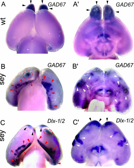
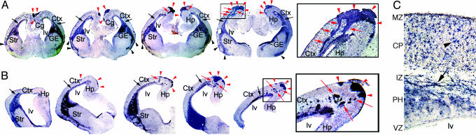
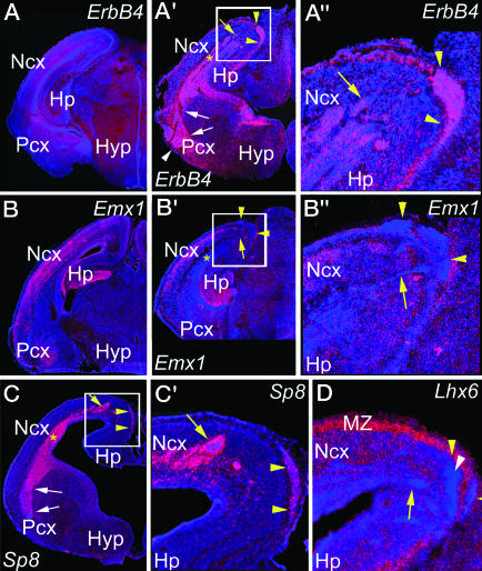
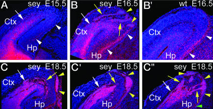
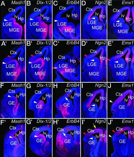
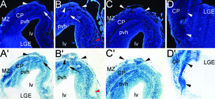
The transcription factor Pax6 is expressed by progenitors in the ventricular zone (VZ) of dorsal telencephalon (dTel), which generate all cortical glutamatergic neurons, but not by progenitors in the medial ganglionic eminence (MGE), which generate cortical GABAergic interneurons (GABA INs), or the lateral ganglionic eminence (LGE), which generate GABA INs that normally migrate to the olfactory bulb. We show that perinatally, Pax6(sey/sey) mice, which lack functional Pax6 protein, have large subpial ectopias in dTel and ventral telencephalon connected by cell streams arising from an aberrant paraventricular ectopia found throughout dTel. The subpial and paraventricular ectopias and connecting cell streams are comprised of postmitotic neurons expressing markers for GABA INs characteristic of a LGE fate. Marker analyses show that dTel VZ progenitors in Pax6 mutants are progressively ventralized, acquiring expression of regulatory genes normally limited to GE progenitors; by midneurogenesis, the entire dTel VZ exhibits ventralization. This ventralization of the dTel VZ is paralleled by the expression of markers for GABA INs superficial to it, suggesting that it ectopically produces GABA INs, leading to their ectopias and a thinner cortical plate due to diminished production of glutamatergic neurons. Genetic lineage tracing demonstrates that the GABA INs comprising the ectopias are from a cortical Emx1 lineage generated in the dTel VZ, definitively showing that dTel progenitors and progeny acquire a ventral, GE, fate in Pax6 mutants. Thus, Pax6 delimits the appropriate proliferative zone for GABA INs and regulates their numbers and distributions by repressing the ventral fates of dTel progenitors and progeny.
Figures
Similar articles
-
Pax6 is required for making specific subpopulations of granule and periglomerular neurons in the olfactory bulb.J Neurosci. 2005 Jul 27;25(30):6997-7003. doi: 10.1523/JNEUROSCI.1435-05.2005. J Neurosci. 2005. PMID: 16049175 Free PMC article.
-
A subpopulation of olfactory bulb GABAergic interneurons is derived from Emx1- and Dlx5/6-expressing progenitors.J Neurosci. 2007 Jun 27;27(26):6878-91. doi: 10.1523/JNEUROSCI.0254-07.2007. J Neurosci. 2007. PMID: 17596436 Free PMC article.
-
Neurotrophin receptor-mediated death of misspecified neurons generated from embryonic stem cells lacking Pax6.Cell Stem Cell. 2007 Nov;1(5):529-40. doi: 10.1016/j.stem.2007.08.011. Cell Stem Cell. 2007. PMID: 18371392
-
Transcriptional Regulation of Cortical Interneuron Development.In: Noebels JL, Avoli M, Rogawski MA, Vezzani A, Delgado-Escueta AV, editors. Jasper's Basic Mechanisms of the Epilepsies. 5th edition. New York: Oxford University Press; 2024. Chapter 47. In: Noebels JL, Avoli M, Rogawski MA, Vezzani A, Delgado-Escueta AV, editors. Jasper's Basic Mechanisms of the Epilepsies. 5th edition. New York: Oxford University Press; 2024. Chapter 47. PMID: 39637134 Free Books & Documents. Review.
-
Transcriptional regulation of olfactory bulb neurogenesis.Anat Rec (Hoboken). 2013 Sep;296(9):1364-82. doi: 10.1002/ar.22733. Epub 2013 Jul 31. Anat Rec (Hoboken). 2013. PMID: 23904336 Review.
Cited by
-
Pax6 mutant cerebral organoids partially recapitulate phenotypes of Pax6 mutant mouse strains.PLoS One. 2022 Nov 28;17(11):e0278147. doi: 10.1371/journal.pone.0278147. eCollection 2022. PLoS One. 2022. PMID: 36441708 Free PMC article.
-
A dlx2- and pax6-dependent transcriptional code for periglomerular neuron specification in the adult olfactory bulb.J Neurosci. 2008 Jun 18;28(25):6439-52. doi: 10.1523/JNEUROSCI.0700-08.2008. J Neurosci. 2008. PMID: 18562615 Free PMC article.
-
The homeobox gene Gsx2 regulates the self-renewal and differentiation of neural stem cells and the cell fate of postnatal progenitors.PLoS One. 2012;7(1):e29799. doi: 10.1371/journal.pone.0029799. Epub 2012 Jan 5. PLoS One. 2012. PMID: 22242181 Free PMC article.
-
A comparative view of human and mouse telencephalon inhibitory neuron development.Development. 2025 Jan 1;152(1):dev204306. doi: 10.1242/dev.204306. Epub 2025 Jan 2. Development. 2025. PMID: 39745314 Review.
-
Altered molecular regionalization and normal thalamocortical connections in cortex-specific Pax6 knock-out mice.J Neurosci. 2008 Aug 27;28(35):8724-34. doi: 10.1523/JNEUROSCI.2565-08.2008. J Neurosci. 2008. PMID: 18753373 Free PMC article.
References
-
- Nadarajah, B. & Parnavelas, J. G. (2002) Nat. Rev. Neurosci. 3, 423-432. - PubMed
-
- Marin, O. & Rubenstein, J. L. R. (2003) Annu. Rev. Neurosci. 26, 441-483. - PubMed
-
- Gonchar, Y. & Burkhalter, A. (1997) Cereb. Cortex 7, 347-358. - PubMed
-
- Anderson, S. A., Eisenstat, D. D., Shi, L. & Rubenstein, J. L. R. (1997) Science 278, 474-476. - PubMed
Publication types
MeSH terms
Substances
Grants and funding
LinkOut - more resources
Full Text Sources
Medical
Molecular Biology Databases
