. 2005 Jun 10;96(11):1169-77.
doi: 10.1161/01.RES.0000169271.33675.05. Epub 2005 May 5.

Functional properties of mouse connexin30.2 expressed in the conduction system of the heart

Affiliations

Functional properties of mouse connexin30.2 expressed in the conduction system of the heart

Maria M Kreuzberg et al. Circ Res. .

Abstract

Gap junction channels composed of connexin (Cx) 40, Cx43, and Cx45 proteins are known to be necessary for impulse propagation through the heart. Here, we report mouse connexin30.2 (mCx30.2) to be a new cardiac connexin that is expressed mainly in the conduction system of the heart. Antibodies raised to the cytoplasmic loop or the C-terminal regions of mCx30.2 recognized this protein in mouse heart as well as in HeLa cells transfected with wild-type mCx30.2 or mCx30.2 fused with enhanced green fluorescent protein (mCx30.2-EGFP). Immunofluorescence analyses of adult hearts yielded positive signals within the sinoatrial node, atrioventricular node, and A-V bundle of the cardiac conduction system. Dye transfer studies demonstrated that mCx30.2 and mCx30.2-EGFP channels discriminate poorly on the basis of charge, but do not allow permeation of tracers >400 Da. Both mCx30.2 and mCx30.2-EGFP gap junctional channels exhibited weak sensitivity to transjunctional voltage (Vj) and a single channel conductance of approximately 9 pS, which is the lowest among all members of the connexin family measured in HeLa cell transfectants. HeLa mCx30.2-EGFP transfectants when paired with cells expressing Cx40, Cx43, or Cx45 formed functional heterotypic gap junction channels that exhibited low unitary conductances (15 to 18 pS), rectifying open channel I-V relations and asymmetric Vj dependence. The electrical properties of homo- and hetero-typic junctions involving mCx30.2 may contribute to slow propagation velocity in nodal tissues and directional asymmetry of excitation spread in the AV nodal region.


Figures
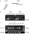
Figure 1
Figure 1
RT-PCR analysis of adult mouse tissues. A, The mCx30.2 gene includes an untranslated exon1 and an exon2 containing the whole coding region (dark shaded) and the 3-untranslated region. The primers (P1-P6) indicated by arrowheads were used for the PCR reaction (for details see Methods in online data supplements). B, Determination of the transcriptional start site was performed with P1-P6 primers and cDNA from heart and brain. C, Intron-spanning RT-PCR of different mouse tissues revealed the expression of the same mCx30.2 transcript isoform in heart, brain, kidney, lung, testis, and very weakly in liver. cDNA of β-actin served as a control.
Figure 2
Figure 2
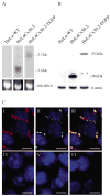
mCx30.2 and mCx30.2-EGFP expression in HeLa transfectants. A, Northern blot analysis of HeLa wild-type, HeLaCx30.2 and HeLaCx30.2-EGFP cells. Ethidium bromide staining of 18S rRNA demonstrates equal loading of total RNA. Blotted RNA was hybridized to a 32P-labeled mCx30.2 probe of the coding region corresponding to the N-terminal portion (nt +1 to nt +530). B, Immunoblot analysis of HeLa-WT, HeLaCx30.2-EGFP and HeLaCx30.2 cells using affinity purified antibodies raised to peptides of the cytoplasmic loop and C-terminal region of mCx30.2. Analysis of β-actin indicates that equal amounts of protein were loaded. C, Immunofluorescence analysis of HeLa cells expressing mCx30.2-EGFP (I-III), Cx40 (IV), Cx43 (V), and Cx45 (VI) with affinity purified antibodies to mCx30.2. Punctate staining of junctional plaques revealed by immunofluorescence (I) and EGFP signals (II) colocalize (III). Cx40, Cx43, and Cx45 do not react with mCx30.2 antibodies (IV-VI). The nuclei of the cells were stained with Hoechst 33528 (blue). Scale bars, 10 μm.
Figure 3
Figure 3
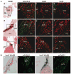
Immunofluorescence analysis of mCx30.2 expression pattern and colocalization with other cardiac connexins. A, Shown are immunofluorescence images (green) of mCx30.2 (b,f,j), Cx40 (c,g,k) and Cx43 (d,h,l) in serial sections from adult mouse heart. Left column shows bright field images (a,e,i) stained for acetylcholine esterase (AChE); squares indicate approximate regions from which immunofluorescence images were acquired. Images in rows (a-d), (e-h) and (i-l) correspond to the regions of the SA node, AV node, and A-V bundle, respectively. Immunostaining of mCx30.2 is visible in the SA node (b), AV node (f), and A-V bundle (j). Cx40 immunostain-ing in the AV node and A-V bundle rarely overlaps with that of mCx30.2, but not within the SA node. Cx43 is not expressed in the SA node, AV node and A-V bundle, but in the atrial and ventricular working myocytes (d,h,l). Dashed lines separate AV-nodal region and A-V bundle from working myocardium. Used abbreviations: AVB, A-V bundle; AVN, AV node; CFB, central fibrous body; IVS, interventricular septum; RA, right atrium; SAN, SA node; SAJ, sinoatrial junction; TV-tricuspid valve. Nuclei of the cells were stained in red with propidium iodide. Scale bars, 20 μm. (B) Coexpression of Cx45 and mCx30.2 in SA node (a,b) and AV node (c,d) of Cx45LacZ mouse. Immunofluorescence signals (b,d; in green) abundantly overlap with LacZ signals (a,c; in blue) in both nodal regions. Scale bars, 20 μm.
Figure 4
Figure 4
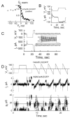
Voltage and chemical gating of mCx30.2 and mCx30.2-EGFP. A, Voltage dependence of mCx30.2 in Xenopus oocytes. Graph of normalized initial and steady-state Gjs (open and filled symbols, respectively) as a function of Vj. Each point represents mean Gj obtained from 11 oocyte pairs. B, Representative junctional currents for Vj steps up to ±120 mV in 20 mV increments. A +20 mV Vj step, 200 ms in duration, preceded each long-duration (30 s) Vj step. C, Gj-Vj plot determined in HeLaCx30.2 (open circles) and HeLaCx30.2-EGFP (filled circles) cell pairs. Solid line is a fit of the data to a regression function of the 5th order. D, Application of heptanol (2 mmol/L) produced full uncoupling of mCx30.2 homotypic junctions within ≈5 s. Recovery was fast and complete. E, Vj and Ij recordings and the resulting gj demonstrating unitary gating events during voltage steps of −105 mV and −125 mV as well as a ramp of -/+100 mV. Single channels were visualized during the recovery from full uncoupling with CO2. Single channel conductance in these recordings was found to be ≈9 pS (see horizontal lines).
Figure 5
Figure 5
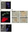
Permeability of mCx30.2, mCx30.2-EGFP, and Cx43 junctions to dyes. A, Phase contrast and fluorescence images demonstrating Alexa Fluor-350 transfer in a HeLaCx30.2-EGFP cell pair; gj=30 nS. Cell 1 was loaded with dye through a patch pipette (left-top). B, Fluorescence images of mCx30.2-EGFP (top; the arrow points to a junctional plaque) and EtBr (bottom) demonstrating EtBr transfer from cell 1 to cell 2; gj=10 nS. C, Phase contrast image overlapping with fluorescence of mCx30.2-EGFP (left; the arrow points to a junctional plaque) and fluorescence image of DAPI (right); gj=6 nS. D, Intercellular transfer of Neurobiotin measured 30 minutes after microinjection into HeLa-WT, HeLa-Cx43, HeLaCx30.2, and HeLaCx30.2-EGFP cells. Neurobiotin does not spread from the injected cell (asterisk) to neighboring cells in a monolayer of HeLa-WT cells, but readily spreads to first and second order neighbors in monolayers of HeLaCx30.2 and HeLaCx30.2-EGFP cells and even more extensively in a monolayer of HeLaCx43 cells. Scale bars, 50 μm.
Figure 6
Figure 6
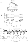
Electrophysiological characterization of heterotypic mCx30.2-EGFP/Cx40 channels examined in cocultured HelaCx30.2-EGFP and HeLaCx40 cells. A, Normalized Gj–Vj plot shows highly asymmetric Vj gating. Data points (open circles) were measured at the ends of the voltage steps. Solid line was obtained applying Vj ramps of 120 s in duration. Vj is defined relative to the mCx30.2-EGFP side. B, Example of Vj, Ij and g traces obtained to voltage steps of 85 and −85 mV applied to the HeLaCx40 cell. Repeated voltage ramps of ±20 mV were used to assess gj between the voltage steps. C, Representative Ij and Vj records measured during application of voltage steps and ramps to the HeLaCx40 cell. Gating transitions to the sub-state (dashed arrows) occurred more often when the mCx30.2 side was made relatively negative. The asterisk shows only one full closure of the channel that occurred at relative negativity of Vj on the Cx40 side.
Figure 7
Figure 7
Gating properties of heterotypic mCx30.2-EGFP/Cx43-CFP channels examined in cell pairs with junctional plaques formed between HeLaCx43-CFP and HeLaCx30.2-EGFP cells. A, Normalized Gj–Vj plot; Vj is defined relative to the mCx30.2-EGFP side. The solid line is a regression line of the fifth order fit to the data. B, Representative Vj and Ij records demonstrate asymmetric Vj gating in response to consecutive steps of −90, 90, −110, and 110 mV and repeated ramps of ±20 mV applied to the HeLaCx30.2-EGFP cell. Negative Vj steps induced a slow decrease in Ij whereas positive ones initially increased and then slowly decreased Ij. C, Single channel record obtained by applying voltage ramps (±105 mV) to the HeLaCx43-CFP cell shows a linear open channel I-V relationship and a single channel conductance of ≈18 pS.
Figure 8
Figure 8
Gating properties of heterotypic mCx30.2-EGFP/Cx45-EGFP channels examined in cocultured HeLaCx30.2-EGFP and N2ACx45-EGFP cells. A, Normalized Gj–Vj plot. The solid line is a fit of the data, shown in solid circles, to the Boltz-mann equation. B,C, Examples of Vj and Ij records demonstrate asymmetric Vj gating in response to voltage steps applied to the Cx45-EGFP side. Repeated voltage ramps of ±20 mV demonstrate reduction of Ij after a positive voltage step (see insert). D, Ij record of single channel in response to ±95 mV voltage steps and ramps (±100 mV) applied to the N2ACx45-EGFP cell. The single open channel I-V relation is essentially linear with a single channel conductance of ≈17 pS (horizontal lines in gj-time plot).

Similar articles

Cited by

References

    1. Söhl G, Willecke K. Gap junctions and the connexin protein family. Cardiovasc Res. 2004;62:228–232. - PubMed
    1. Bukauskas FF, Verselis VK. Gap junction channel gating. Biochim Biophys Acta. 2004;1662:42–60. - PMC - PubMed
    1. Gros DB, Jongsma HJ. Connexins in mammalian heart function. Bioessays. 1996;18:719–730. - PubMed
    1. Severs NJ, Dupont E, Coppen SR, Halliday D, Inett E, Baylis D, Rothery S. Remodelling of gap junctions and connexin expression in heart disease. Biochim Biophys Acta. 2004;1662:138–148. - PubMed
    1. Lo CW. Role of gap junctions in cardiac conduction and development: insights from the connexin knockout mice. Circ Res. 2000;87:346–348. comment] [editorial. - PubMed

Publication types