

doi: 10.1529/biophysj.105.064816.
Epub 2005 May 6.
Cholesterol-enriched lipid domains can be visualized by di-4-ANEPPDHQ with linear and nonlinear optics
Affiliations
- PMID: 15879475
- PMCID: PMC1366585
- DOI: 10.1529/biophysj.105.064816
Item in Clipboard
Cholesterol-enriched lipid domains can be visualized by di-4-ANEPPDHQ with linear and nonlinear optics
Biophys J.
2005 Jul.
Abstract
We present a membrane-staining dye, di-4-ANEPPDHQ, which differentiates liquid-ordered phases from liquid-disordered phases coexisting in model membranes under both linear and nonlinear microscopies. The dye's fluorescence emission spectrum is blue-shifted 60 nm in liquid-ordered phases compared with liquid-disordered phases, and shows strong second harmonic generation in the liquid-disordered phase compared with the liquid-ordered phase. The ease of staining and the ability of this single dye to detect both phases, should lead to broad applications in biophysical studies of lipid domains in model membranes and cells.
Figures
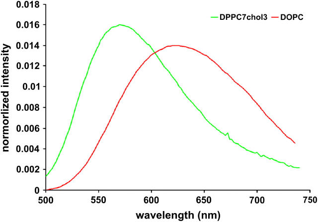
Emission spectra of di-4-ANEPPDHQ in LUVs of pure DOPC (red) and LUVs of 7:3 DPPC/cholesterol (green).
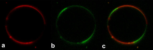
Confocal fluorescence images of a GUV (2:2:1 DPPC/DOPC/cholesterol) stained with 1 μM di-4-ANEPPDHQ. (a) Long-pass 650-nm channel; (b) band-pass 500–530-nm channel; (c) merge of panels a and b.
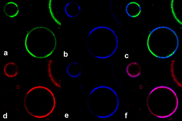
TPF (red and green) and SHG (blue) images of GUVs with both phases stained with di-4-ANEPPDHQ. (a) TPF from the 515–565-nm emission channel; (b) SHG signal taken simultaneously with a; (c) merge of a and b; (d) TPF from the 650–700-nm emission channel; (e) SHG signal taken simultaneously with d; (f) merge of d and e.
References
-
- Brown, D., and J. Rose. 1992. Sorting of GPI-anchored proteins to glycolipid-enriched membrane subdomains during transport to the apical cell surface. Cell. 68:533–544. - PubMed
-
- Bagatolli, L. A., S. A. Sanchez, T. Hazlett, and E. Gratton. 2003. Giant vesicles, laurdan, and two-photon fluorescence microscopy: evidence of lipid lateral separation in bilayers. Methods Enzymol. 360:481–500. - PubMed
Publication types
MeSH terms
Substances
Grants and funding
LinkOut - more resources
Full Text Sources
Other Literature Sources
Medical
