. 2005 May;4(5):911-9.
doi: 10.1128/EC.4.5.911-919.2005.

Cell biology of conidial anastomosis tubes in Neurospora crassa

Affiliations

Cell biology of conidial anastomosis tubes in Neurospora crassa

M Gabriela Roca et al. Eukaryot Cell. 2005 May.

Abstract

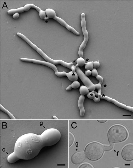
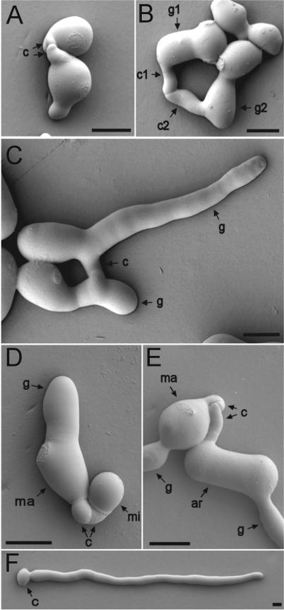
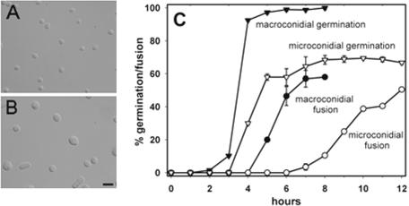
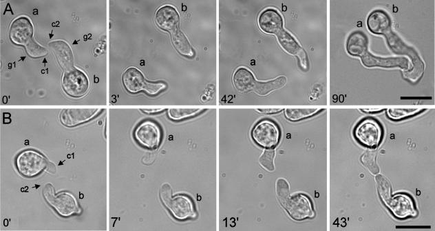
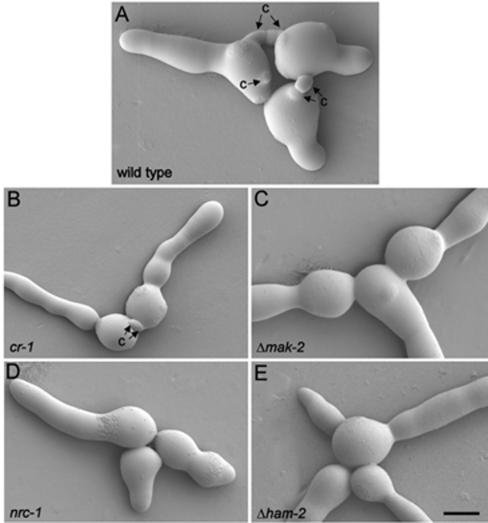
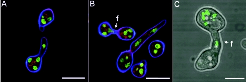
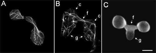
Although hyphal fusion has been well documented in mature colonies of filamentous fungi, it has been little studied during colony establishment. Here we show that specialized hyphae, called conidial anastomosis tubes (CATs), are produced by all types of conidia and by conidial germ tubes of Neurospora crassa. The CAT is shown to be a cellular element that is morphologically and physiologically distinct from a germ tube and under separate genetic control. In contrast to germ tubes, CATs are thinner, shorter, lack branches, exhibit determinate growth, and home toward each other. Evidence for an extracellular CAT inducer derived from conidia was obtained because CAT formation was reduced at low conidial concentrations. A cr-1 mutant lacking cyclic AMP (cAMP) produced CATs, indicating that the inducer is not cAMP. Evidence that the transduction of the CAT inducer signal involves a putative transmembrane protein (HAM-2) and the MAK-2 and NRC-1 proteins of a mitogen-activated protein kinase signaling pathway was obtained because ham-2, mak-2, and nrc-1 mutants lacked CATs. Optical tweezers were used in a novel experimental assay to micromanipulate whole conidia and germlings to analyze chemoattraction between CATs during homing. Strains of the same and opposite mating type were shown to home toward each other. The cr-1 mutant also underwent normal homing, indicating that cAMP is not the chemoattractant. ham-2, mak-2, and nrc-1 macroconidia did not attract CATs of the wild type. Fusion between CATs of opposite mating types was partially inhibited, providing evidence of non-self-recognition prior to fusion. Microtubules and nuclei passed through fused CATs.


Figures

FIG. 1.
FIG. 1.
CATs and germ tubes. (A) Germinated macroconidia with long germ tubes avoiding each other and CATs which have grown (homed) toward each other and fused (asterisks). The image was obtained by low-temperature scanning electron microscopy. Bar, 10 μm. (B) A single macroconidium which has produced a germ tube (g) and a CAT (c). The image was obtained by low-temperature scanning electron microscopy. Bar, 2.5 μm. (C) Fusion of CATs (f) produced directly from two conidia, one of which has produced a germ tube (g). This is a differential interference contrast image. Bar, 2.5 μm.
FIG. 2.
FIG. 2.
(A to C) Different types of CATs. (A) CATs (c) produced directly from macroconidia. (B) A CAT (c1) produced directly from a germ tube tip (g1) and a CAT (c2) produced just behind a germ tube tip (g2). Note that the germ tubes have avoided each other, while the CATs have homed toward each other, are narrower, and have fused. (C) Fused CATs (c) originally produced as side branches of germ tubes (g). (D) CATs (c) produced by a macroconidium (ma) fused with one produced by a microconidium (mi). Note that the germ tube (g) produced by the macroconidium is much wider than the CATs. (E) CATs (c) produced by a macroconidium (ma) fused with one produced by an arthroconidium (ar). Both conidia have also produced wider germ tubes (g). (F) Solitary macroconidium (c) which has produced a germ tube but not a CAT (cf. with Fig. 1B). The conidial concentration in this preparation was lower (105 conidia per ml) than normal (106 conidia per ml). Images were obtained by low-temperature scanning electron microscopy. Bars, 5 μm.
FIG. 3.
FIG. 3.
Microconidial and macroconidial germination and fusion after purification by FACS. (A) Sample of predominately microconidia which were <4 μm in length. (B) Sample of predominately macroconidia that were 7 to 12 μm in length. Bar, 25 μm. (C) Time courses of germination and fusion of microconidia and macroconidia purified by FACS.
FIG. 4.
FIG. 4.
Optical tweezer homing assay experiments. (A) Control experiment. At 0 min, CATs (c1 and c2) formed from the thicker tips of germ tubes (g1 and g2) of two macroconidia (a and b) are homing toward each other. At 3 min, the two germlings have been moved apart with the optical tweezers. At 42 min, the CAT tips are reorientating their growth back toward each other. At 90 min, the CAT tips have adhered and fused with each other. (B) CATs (c1 and c2) formed directly from conidia (a and b) of a cr-1 strain lacking cAMP home toward each other. At 0 min, the CATs are homing toward each other. At 7 min, the two CATs have been moved apart with optical tweezers. At 13 min, the CAT tips have reorientated their growth back toward each other. At 43 min, the CATs have adhered and fused. Bars, 10 μm.
FIG. 5.
FIG. 5.
MAP kinase mutants defective in CAT formation. (A) Wild-type strain with CATs (c); (B) cr-1 mutant lacking cAMP produces CATs; (C) Δmak-2 mutant lacking CATs; (D) nrc-1 mutant lacking CATs; (E) ham-2 mutant lacking CATs. Images were obtained by low-temperature scanning electron microscopy. Bar, 5 μm.
FIG. 6.
FIG. 6.
Nuclear migration and CAT fusion between macroconidia. (A) Homing CATs between macroconidia of a mat a strain. Note that a nucleus has migrated to a region just behind the tip in the lower CAT. Nuclei are labeled with H1-GFP (green); membranes are labeled with FM4-64 (red); cell walls are labeled with calcofluor white (blue). The image was obtained by confocal microscopy. Bar, 10 μm. (B) Fused CATs (f) between macroconidia of a mat a strain. Note that a nucleus is migrating through the fused CATs. Staining is as described for panel A; the image was obtained by confocal microscopy. Bar, 10 μm. (C) Fused CATs (f) between macroconidia: one (labeled with nuclear-targeted H1-GFP) is of a mat a strain and the other is of a mat A strain. Nuclei are fluorescing green. This is a merged confocal image and a transmitted bright-field image. Bar, 5 μm.
FIG. 7.
FIG. 7.
(A) Microtubules (labeled with β-tubulin-GFP) longitudinally organized and extending towards CAT tips of macroconidia which are homing toward each other. (B) After fusion, microtubules (labeled with β-tubulin-GFP) have extended through the fused CATs (f) from one macroconidium (c) bearing a germ tube (g) into the other and vice versa and, as a result, have become intermixed. (C) Germ tube (g) which is growing out of fused CATs. Images were obtained by confocal microscopy. Bar, 5 μm.

References

    1. Ashkin, A., J. M. Dziedzic, and T. Yamane. 1987. Optical trapping and manipulation of single cells using infrared laser beams. Nature 330:769-771. - PubMed
    1. Aubrey, L., and R. Firtel. 1999. Integration of signalling networks that regulate Dictyostelium differentiation. Annu. Rev. Cell Dev. Biol. 15:469-517. - PubMed
    1. Bistis, G. N. 1981. Chemotropic interactions between trichogynes and conidia of opposite mating-type in Neurospora crassa. Mycologia 73:959-975.
    1. Bobrovicz, P., R. Pawlak A. Corea, D. Bell-Pedersen, and D. J. Ebbole. 2002. The Neurospora crassa pheromone precursor genes are regulated by the mating type locus and the circadian clock. Mol. Microbiol. 45:795-804. - PubMed
    1. Bölker, M., M. Urban, and R. Kahmann. 1992. The a mating type locus of U. maydis specified cell signaling components. Cell 68:441-450. - PubMed

Publication types

MeSH terms

LinkOut - more resources