

The CD154-CD40 T-cell co-stimulation pathway in liver ischemia and reperfusion inflammatory responses
- PMID: 15880047
- PMCID: PMC4470618
- DOI: 10.1097/01.tp.0000161248.43481.a2
The CD154-CD40 T-cell co-stimulation pathway in liver ischemia and reperfusion inflammatory responses
Abstract
Background: Ischemia-reperfusion (I/R) injury is a prime antigen-independent inflammatory factor in the dysfunction of liver transplants. The precise contribution of T cells in the mechanism of I/R injury remains to be elucidated. As the CD154-CD40 co-stimulation pathway provides essential second signal in the initiation and maintenance of T-cell-dependent immune responses, this study was designed to assess the role of CD154 signaling in the pathophysiology of liver I/R injury.
Methods: A mouse model of partial 90-min warm hepatic ischemia followed by 6 hr of reperfusion was used. Three animal groups were studied: (1) wild-type (WT) mice treated with Ad-(-gal versus Ad-CD40 immunoglobulin; (2) untreated WT versus CD154 (MR1) monoclonal antibody-treated WT mice; and (3) untreated WT versus CD154 knockout mice.
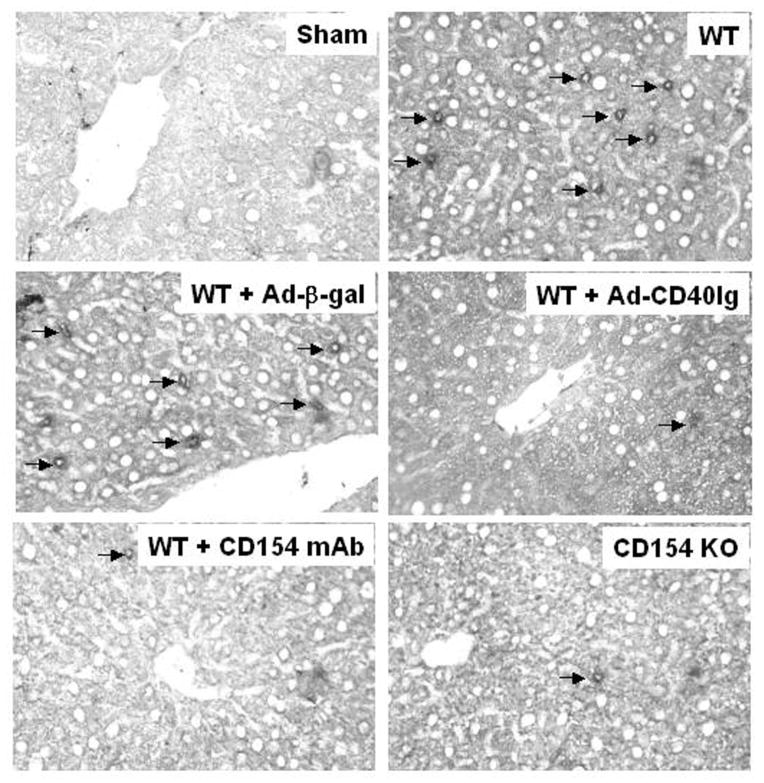
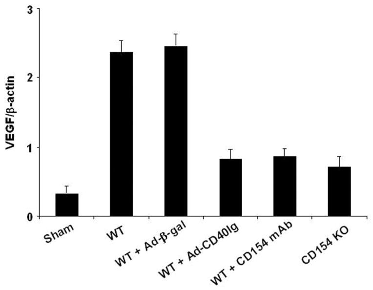
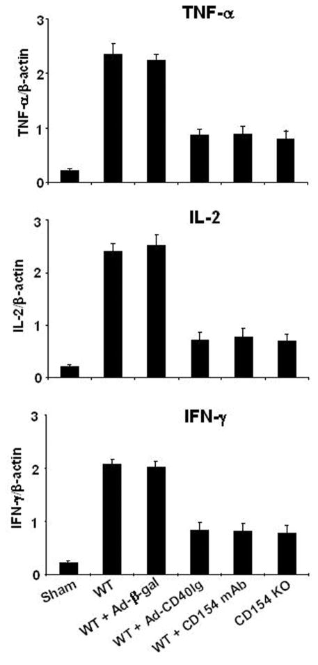
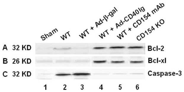
Results: The disruption of CD154 signaling in all three animal groups ameliorated otherwise fulminant liver injury, as evidenced by depressed serum glutamic oxaloacetic transaminase levels, compared with controls. These beneficial effects were accompanied by depressed hepatic T-cell sequestration, local decrease of vascular endothelial growth factor expression, inhibition of tumor necrosis factor-(and T-helper type 1 cytokine production, and induction of antiapoptotic (Bcl-2/Bcl-xl) but depression of proapoptotic (caspase-3) proteins.
Conclusions: By using in parallel a gene therapy approach, pharmacologic blockade, and genetically targeted mice, these findings document the benefits of disrupting CD154 to selectively modulate inflammatory responses in liver I/R injury. This study reinforces the key role of CD154-CD40 T-cell co-stimulation in the pathophysiology of liver I/R injury.
Figures

Similar articles
-
CD154-CD40 T-cell costimulation pathway is required in the mechanism of hepatic ischemia/reperfusion injury, and its blockade facilitates and depends on heme oxygenase-1 mediated cytoprotection.Transplantation. 2002 Aug 15;74(3):315-9. doi: 10.1097/00007890-200208150-00005. Transplantation. 2002. PMID: 12177608
-
Gene therapy for liver transplantation using adenoviral vectors: CD40-CD154 blockade by gene transfer of CD40Ig protects rat livers from cold ischemia and reperfusion injury.Mol Ther. 2004 Jan;9(1):38-45. doi: 10.1016/j.ymthe.2003.10.011. Mol Ther. 2004. PMID: 14741776 Free PMC article.
-
CD4 T cells promote tissue inflammation via CD40 signaling without de novo activation in a murine model of liver ischemia/reperfusion injury.Hepatology. 2009 Nov;50(5):1537-46. doi: 10.1002/hep.23153. Hepatology. 2009. PMID: 19670423 Free PMC article.
-
The role of CD40-CD154 interaction in cell immunoregulation.J Biomed Sci. 2004 Jul-Aug;11(4):426-38. doi: 10.1007/BF02256091. J Biomed Sci. 2004. PMID: 15153777 Review.
-
The CD40/CD154 receptor/ligand dyad.Cell Mol Life Sci. 2001 Jan;58(1):4-43. doi: 10.1007/pl00000776. Cell Mol Life Sci. 2001. PMID: 11229815 Free PMC article. Review.
Cited by
-
New frontiers for platelet CD154.Exp Hematol Oncol. 2015 Mar 1;4:6. doi: 10.1186/s40164-015-0001-6. eCollection 2015. Exp Hematol Oncol. 2015. PMID: 25763299 Free PMC article.
-
Activation of CD40 with platelet derived CD154 promotes reactive oxygen species dependent death of human hepatocytes during hypoxia and reoxygenation.PLoS One. 2012;7(1):e30867. doi: 10.1371/journal.pone.0030867. Epub 2012 Jan 25. PLoS One. 2012. PMID: 22295117 Free PMC article.
-
AICAR-Induced AMPK Activation Inhibits the Noncanonical NF-κB Pathway to Attenuate Liver Injury and Fibrosis in BDL Rats.Can J Gastroenterol Hepatol. 2018 Dec 19;2018:6181432. doi: 10.1155/2018/6181432. eCollection 2018. Can J Gastroenterol Hepatol. 2018. PMID: 30662889 Free PMC article.
-
Lymphocytes and ischemia-reperfusion injury.Transplant Rev (Orlando). 2009 Jan;23(1):1-10. doi: 10.1016/j.trre.2008.08.003. Transplant Rev (Orlando). 2009. PMID: 19027612 Free PMC article. Review.
-
Pharmacological inhibition of MyD88 homodimerization counteracts renal ischemia reperfusion-induced progressive renal injury in vivo and in vitro.Sci Rep. 2016 Jun 1;6:26954. doi: 10.1038/srep26954. Sci Rep. 2016. PMID: 27246399 Free PMC article.
References
-
- Farmer DG, Amersi F, Kupiec-Weglinski JW, et al. Current status of ischemia and reperfusion injury in the liver. Transplant Rev. 2000;14:106.
-
- Fondevila C, Busuttil RW, Kupiec-Weglinski JW. Hepatic ischemia/reperfusion injury: A fresh look. Exp Mol Pathol. 2003;74:86. - PubMed
-
- Bulkley GB, Oshima A, Bailey RW. Pathophysiology of hepatic ischemia in cardiogenic shock. Am J Surg. 1996;151:87. - PubMed
Publication types
MeSH terms
Substances
Grants and funding
LinkOut - more resources
Full Text Sources
Research Materials
