. 2005 Jun;11(6):678-82.
doi: 10.1038/nm1247. Epub 2005 May 8.

Quantum dots spectrally distinguish multiple species within the tumor milieu in vivo

Affiliations

Quantum dots spectrally distinguish multiple species within the tumor milieu in vivo

Mark Stroh et al. Nat Med. 2005 Jun.

Abstract

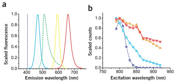
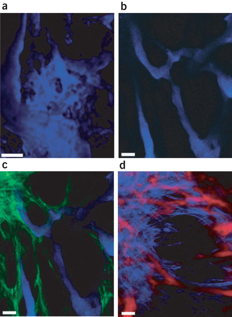
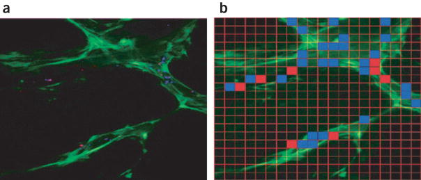
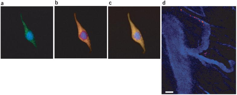
A solid tumor is an organ composed of cancer and host cells embedded in an extracellular matrix and nourished by blood vessels. A prerequisite to understanding tumor pathophysiology is the ability to distinguish and monitor each component in dynamic studies. Standard fluorophores hamper simultaneous intravital imaging of these components. Here, we used multiphoton microscopy techniques and transgenic mice that expressed green fluorescent protein, and combined them with the use of quantum dot preparations. We show that these fluorescent semiconductor nanocrystals can be customized to concurrently image and differentiate tumor vessels from both the perivascular cells and the matrix. Moreover, we used them to measure the ability of particles of different sizes to access the tumor. Finally, we successfully monitored the recruitment of quantum dot-labeled bone marrow-derived precursor cells to the tumor vasculature. These examples show the versatility of quantum dots for studying tumor pathophysiology and creating avenues for treatment.


Conflict of interest statement

COMPETING INTERESTS STATEMENT

The authors declare that they have no competing financial interests.

Figures

Figure 1
Figure 1
Spectra corresponding to representative quantum dots used in this study. (a) The family of emission spectra was generated from quantum dots tuned to emit at wavelengths not shared by signals from GFP and second harmonic generation (green and dark blue dashed lines, respectively). In this depiction, second harmonic generation is generated with 810-nm incident light. The quantum dot emission spectra were narrow and symmetric; corresponding emission peaks and associated full-width half-maximum values were 470 and 30, 590 and 30, and 660 and 40 nm for QD470 (light blue line), QD590 (orange line) and QD660 (red line), respectively. (b) The relative two-photon action cross-sections (scaled to a maximum value of 1) for the corresponding quantum dot and cascade blue (dark blue line). Owing to the relatively broad two-photon action cross sections, the quantum dot may be excited concomitantly with the intrinsic second harmonic generation and GFP sources using multiphoton microscopy.
Figure 2
Figure 2
Vascular imaging with quantum dots. (a) The depth projection of 20 images taken at 5-μm intervals depicts pockets of labeled dextrans that have extravasated and collected in the interstitium, resulting in blurred tumor vessels. (b) When QD470 is used as a vessel marker, the depth projection of 15 images obtained at 5-μm intervals shows a sharp boundary between vessel and interstitium. (c) Concurrent imaging of both QD470 and GFP (indicated in green) provides clear separation of vessel from GFP-expressing perivascular cells. (d) Vessels highlighted with a deep red (QD660) micelle preparation were imaged simultaneously with the SHG signal (indicated in blue); the image represents a projection of a stack of 20 images at an interval of 2 μm per slice. Scale bar, 50 μm.
Figure 3
Figure 3
Quantum dot–loaded beads as drug delivery vehicle. Both 100 and 500 nm silica microspheres were loaded with QD470 and QD590, respectively, and administered to a VEGF-GFP mouse bearing a MCaIV tumor explant in a skinfold chamber. (a) After a 5-h period, a depth projection of five images taken at 5-μm intervals showed a heterogeneous extravasation pattern. (b) This image was meshed with a 20 × 20 grid, and regions that correspond to a maximum extravasated particle size of 500 nm and 100 nm are indicated with red and blue grids, respectively. In this fashion, we assessed the ability of candidates to access VEGF-GFP perivascular cells. The image is 707 μm across.
Figure 4
Figure 4
Cell labeling and tracking using quantum dot–TAT bioconjugate. (ac) Three depictions of a mouse endothelial cell loaded with QD590-TAT and incubated with CellTracker CMFDA as a control. (a) CellTracker CMFDA signal (green). (b) QD590-TAT signal (orange). (c) Colocalization of signals in a and b. In ac, blue corresponds to the DAPI nuclear stain. (d) Seven images superimposed in time as a single bone marrow lineage-negative cell labeled with QD590-TAT (orange) navigated the tumor vessels highlighted with QD470 micelles (blue) in vivo. For ac, the image was obtained using a ×63, 1.2 NA water-immersion lens, and represents the depth projection of 41 images taken at 0.75-μm intervals. The images are 112 μm across. For d, the image represents seven repeated scans at a fixed depth (~100 μm) taken at 1-s intervals. Scale bar, 50 μm.

References

    1. Jain RK, Munn LL, Fukumura D. Dissecting tumour pathophysiology using intravital microscopy. Nat Rev Cancer. 2002;2:266–276. - PubMed
    1. Brown EB, et al. In vivo measurement of gene expression, angiogenesis and physiological function in tumors using multiphoton laser scanning microscopy. Nat Med. 2001;7:864–868. - PubMed
    1. Fukumura D, et al. Tumor induction of VEGF promoter activity in stromal cells. Cell. 1998;94:715–725. - PubMed
    1. Zipfel WR, Williams RM, Webb WW. Nonlinear magic: multiphoton microscopy in the biosciences. Nat Biotechnol. 2003;21:1369–1377. - PubMed
    1. Brown E, et al. Dynamic imaging of collagen and its modulation in tumors in vivo using second-harmonic generation. Nat Med. 2003;9:796–800. - PubMed

Publication types

MeSH terms

Substances